Root Canal Treatment
Will the Tooth Be Dead
After a Root Canal?
No. A root canal removes the pulp from inside the tooth, but the tooth itself remains alive at the bone interface via the periodontal ligament. Survival rates exceed 95 percent at 5 years and 70 percent at 30 years when properly crowned.

The short version
- The "dead tooth" phrase is shorthand at best, wrong at worst. The pulp inside the tooth is removed. The periodontal ligament outside the root remains intact with its own independent blood supply and nerve supply.
- The periodontal ligament keeps the tooth anchored, nourished, and pressure-sensing. You still feel biting force normally after a root canal because the PDL mechanoreceptors are undisturbed.
- Survival rates are strong over decades: 95 percent at 5 years, 85 to 90 percent at 10 years, 80 to 85 percent at 20 years, 70 to 75 percent at 30 years with a crown and routine maintenance.
- What changes is internal: no hot/cold sensitivity, gradually more brittle dentin, possible slow darkening, no pulp pain to warn you about new decay. Crown, checkups, and hygiene address all three.
Two life-support systems inside one tooth
The confusion about whether a root canal tooth is dead comes from treating the tooth as a single structure with a single life-support system. In reality, a tooth has two distinct biological systems, and a root canal removes only one of them.
The first is the pulp: the soft tissue chamber running through the centre of the crown and down into the root canals. The pulp contains blood vessels and nerve fibres that nourish the dentin from within and carry pain signals. When the pulp becomes infected or necrotic, root canal treatment removes it.
The second is the periodontal ligament (PDL): a thin layer of fibrous connective tissue sitting in the 0.15 to 0.38 millimetre gap between the root surface and the surrounding bone socket. The PDL has its own independent blood supply, its own nerve fibres, and its own population of cells including fibroblasts, osteoblasts, cementoblasts, and immune cells. A root canal procedure does not enter this space. The PDL is completely undisturbed.

The clearest way to describe a root canal tooth is with the phrase used in modern endodontic teaching: non-vital pulp, vital tooth. The pulp tissue is gone. The tooth as a functioning unit of the dental arch is alive at the bone interface and acts like a normal tooth for almost every practical purpose.
What the periodontal ligament does after the pulp is removed
Understanding the PDL's role after root canal treatment is what makes the "not dead" claim concrete rather than reassuring but vague.
- Mechanical anchoring through Sharpey fibres The PDL is made of collagen fibre bundles, called Sharpey fibres, that run from the cementum on the outer root surface into the surrounding alveolar bone. These fibres run at different angles in different zones of the root to resist both vertical chewing force and lateral movement. When you bite down, the tooth depresses very slightly in the socket and the PDL springs it back. This slight give protects both the tooth and the bone from impact damage.
- Three independent blood sources, none from the pulp The PDL receives blood from the apical artery branch (reaching the PDL through an exit separate from the pulp canal), from the gingival tissue above, and from the alveolar bone itself. When the pulp blood supply is removed during root canal treatment, the gingival and alveolar sources continue without interruption. The PDL is never deprived of perfusion by the procedure.
- Pressure sensation fully intact The PDL contains highly tuned mechanoreceptors that detect the direction and force of bite. This is why you can feel a small seed between your teeth without pressing too hard, and why you instantly know if a piece of bone is in your food. These receptors are in the PDL, not the pulp. After a root canal, they function normally. You feel pressure on the tooth. What you lose is temperature sensitivity (hot and cold), which was a pulp function, not a PDL function.
- Active bone remodelling continues The PDL transmits mechanical signals to the surrounding bone. Osteoblasts and osteoclasts in the socket respond to these signals and remodel the bone over time. This is what makes orthodontic tooth movement possible on root canal-treated teeth. The PDL never stops working, and the bone around the tooth remains metabolically active and healthy as long as the PDL is intact.
- Immune access to the root surface Immune cells reach the area around the root tip through the PDL blood supply. If a small residual infection is present after a root canal, the immune system can still mount a localised defence at the apex through the PDL route. This is why minor periapical infections often resolve on their own after the infected pulp has been removed and the canal sealed.

The 30-year survival numbers
Survival data from large endodontic outcome studies and follow-up at Picasso provide the clearest answer to whether root canal teeth are "dead." Dead structures do not survive at 95 percent for five years and 70 percent for thirty. They wear out. They fracture. They develop secondary problems the same way any restored tooth does over decades. But they function.
With crown and maintenance
- 5 years: 95 percent survival
- 10 years: 85 to 90 percent survival
- 20 years: 80 to 85 percent survival
- 30 years: 70 to 75 percent survival
- 40+ years: 55 to 65 percent (limited data)
Without crown (posterior teeth)
- 5 years: 70 to 85 percent survival
- 10 years: 55 to 70 percent survival
- 20 years: 35 to 50 percent survival
- 30 years: 20 to 30 percent survival
- 40+ years: 10 to 20 percent survival
The survival gap between crowned and uncrowned root canal teeth is almost entirely explained by vertical root fracture. Dehydrated dentin develops microcracks under decades of chewing force. Without a crown distributing those forces, the cracks propagate until the root splits and the tooth cannot be saved. With a crown, the same dehydrated dentin is protected and the fracture risk drops from 15 to 30 percent over five years to 1 to 2 percent. See the full analysis in why a crown is needed after root canal treatment.
Had a root canal some years ago and wondering whether the tooth is still in good shape? A review appointment at Picasso Dental includes X-ray assessment of the periapical area and PDL space, and a check of the crown seal integrity.
Book a reviewWhat genuinely changes after the pulp is removed
The tooth is not dead, but it is also not unchanged. The full honest picture requires naming both what stays the same and what shifts.
What stays the same
- Tooth anchored in jaw via PDL
- Pressure sensation when biting (PDL mechanoreceptors)
- Bone around tooth remains stimulated
- Can chew with full force (with crown)
- Can still be moved orthodontically
- Immune access to root surface intact
- Cementum on root stays nourished
- No implant or bridge needed
What changes
- No hot/cold temperature sensitivity
- No electrical pulp test response
- Cavity warning system reduced (no pulp pain)
- Dentin becomes gradually more brittle
- Possible slow grey or yellow darkening
- Crown required for posterior teeth
- 6-monthly checkups more important than before
- Decay at crown margin is silent without pulp
The loss of temperature sensitivity is actually the change that most affects long-term management. A vital tooth uses hot and cold pain as an early-warning system for new decay. A root canal tooth cannot do this. This is why 6-monthly X-ray and clinical recall is more important for root canal-treated teeth than for vital ones. Decay can develop around the crown margin without any symptoms, and by the time it becomes visible it may have compromised the tooth beyond repair.
"I performed a root canal and they placed a porcelain crown to protect the tooth. I have been pain free since. The process was easy and the prices were as quoted to me, no surprises."
Garrett Lee, United States, Hanoi Old Quarter, Google review
How root canal teeth eventually fail when they do
Long-term root canal tooth failure is almost always mechanical or hygienic, not biological. Patients ask whether the tooth died. The answer is usually no: the tooth wore out or developed a secondary problem at one of three predictable points.
- Vertical root fracture The most common failure mode, especially in posterior teeth without a crown. The access cavity and canal shaping remove significant dentin. What remains is thinner and, over years without internal hydration from the pulp, gradually more brittle. Chewing force creates microcracks. A crack that propagates the length of the root makes the tooth unrestorable and requires extraction. Crowning within the 2 to 4 week window after the root canal reduces this risk by an order of magnitude.
- Recurrent decay at the crown margin If the seal between crown and tooth begins to leak, bacteria enter the underlying tooth and cause new decay. Because there is no pulp, there is no pain to signal this is happening. Detection requires the 6-monthly clinical examination and X-ray. Without recall, crown-margin decay often reaches an advanced stage before it becomes visible or symptomatic, sometimes deep enough to remove too much tooth structure for a new crown to hold.
- Gum disease loosening the PDL attachment Chronic periodontitis resorbs bone and detaches the PDL fibre from the socket around any tooth, root canal-treated or not. Root canal teeth are not more susceptible to periodontitis than vital teeth. But because they lack the pulp pain warning, patients with chronic gum disease and a root canal tooth can sometimes reach a more advanced periodontal stage before seeking treatment. Regular hygiene visits are the preventive measure.

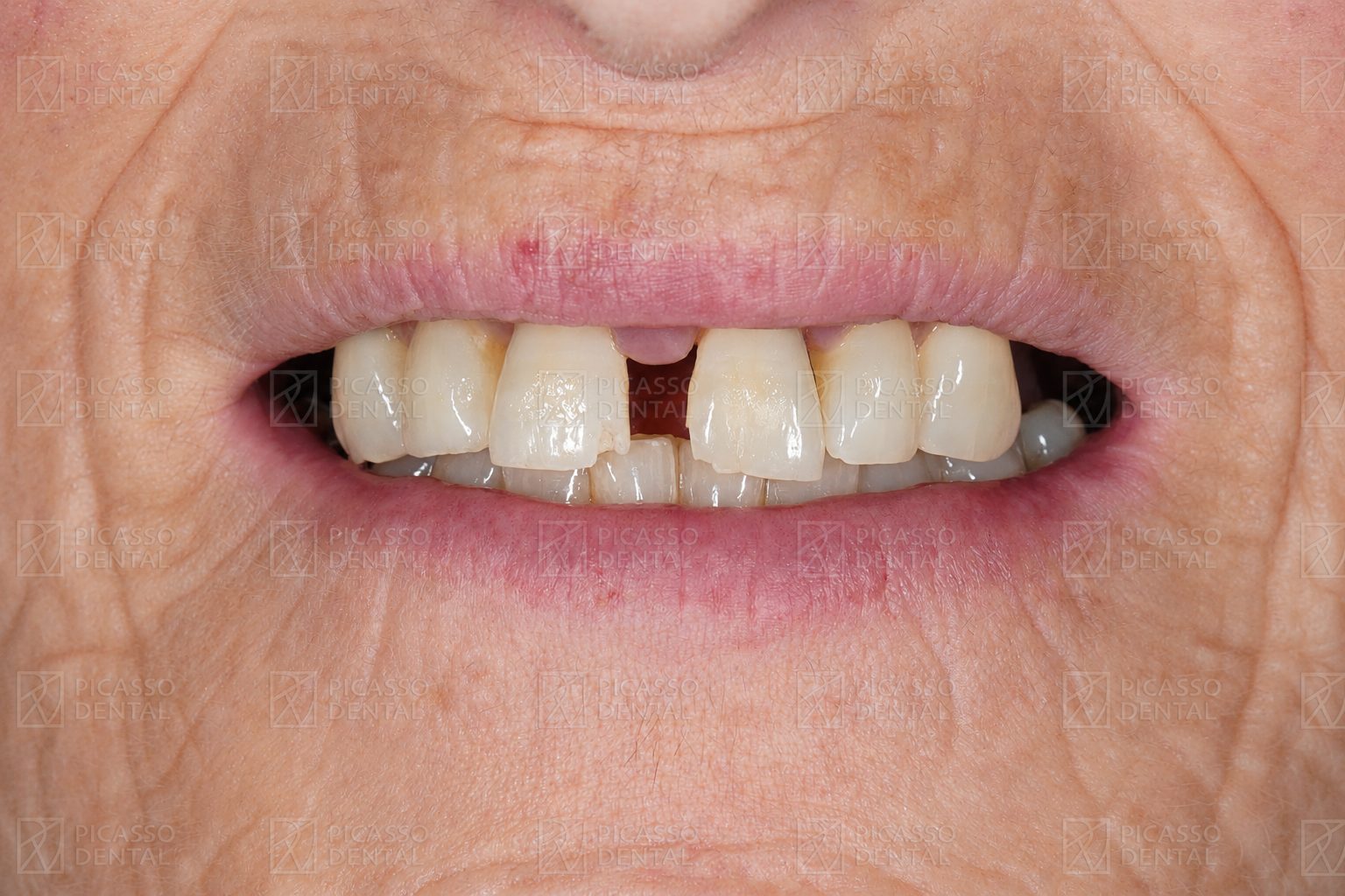 Before
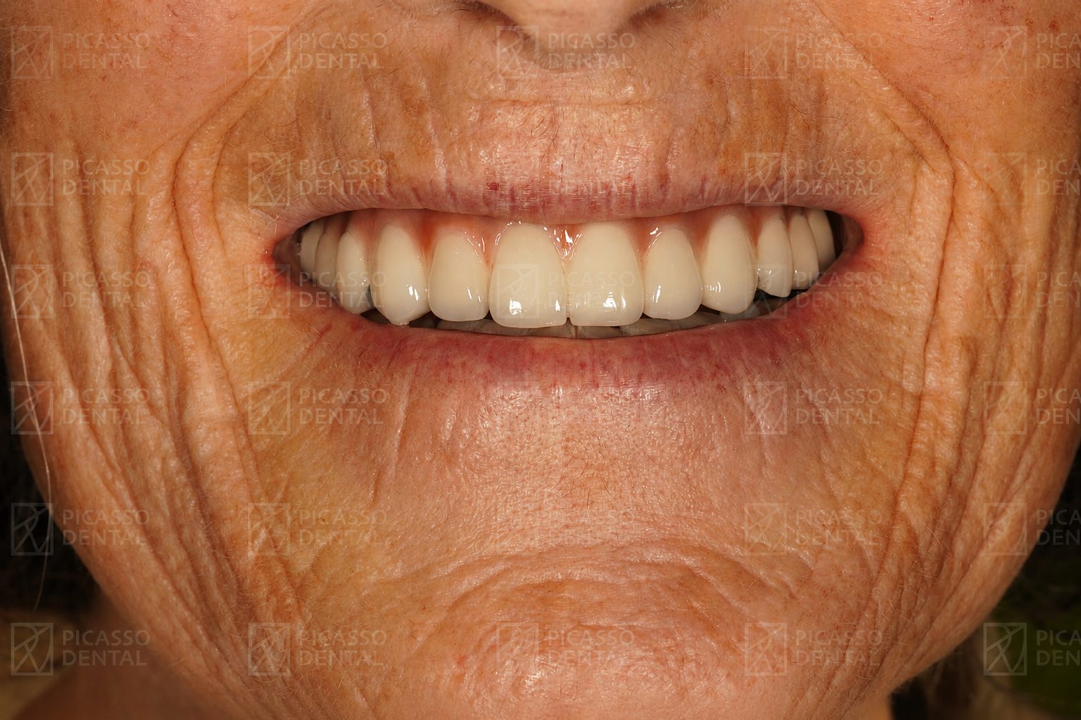 After
AfterRoot canal and zirconia crown. Tooth functional and PDL intact.
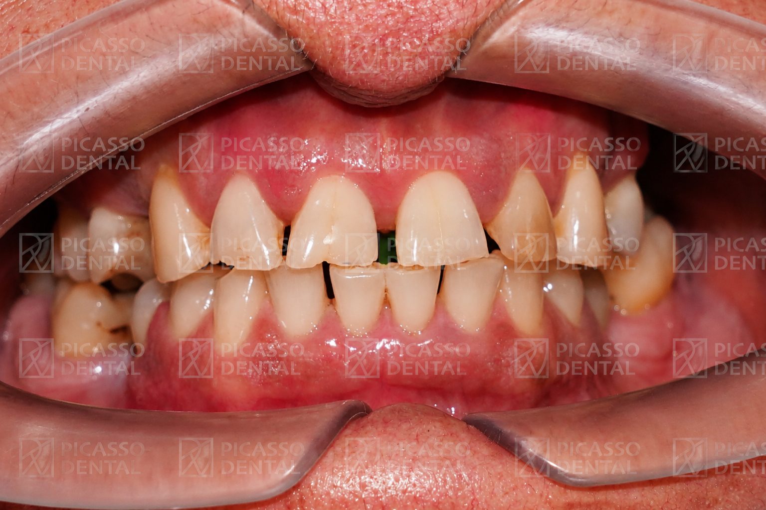 Before
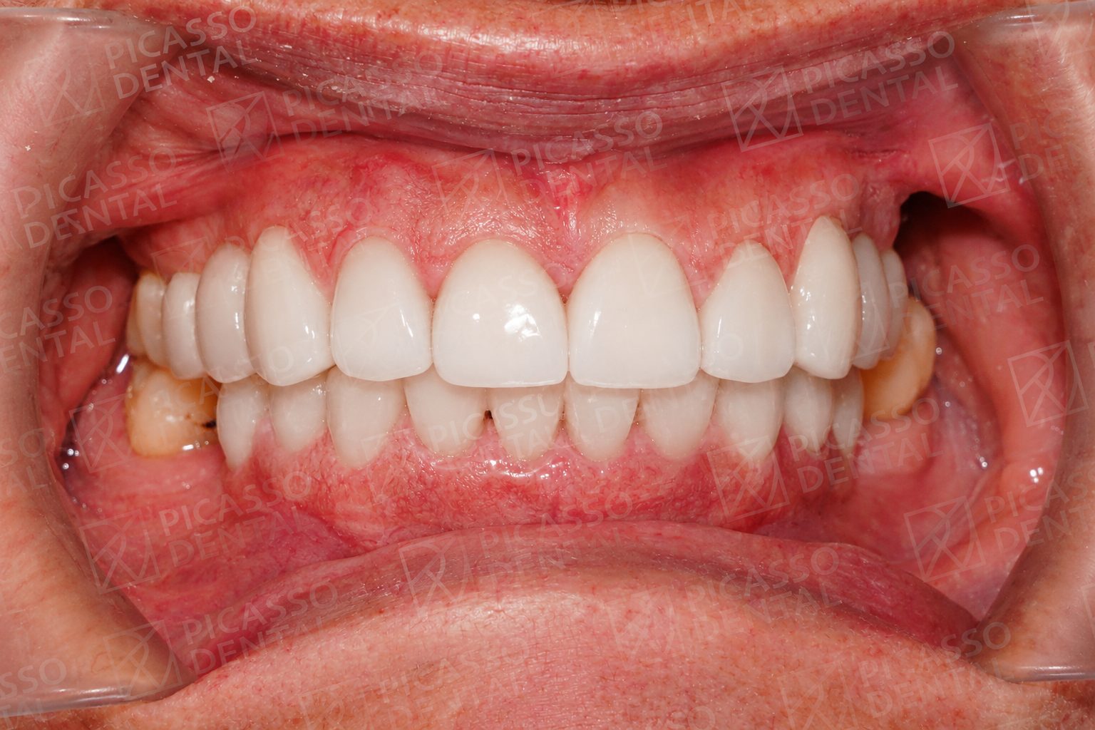 After
AfterRoot canal followed by porcelain crown within 2 weeks.
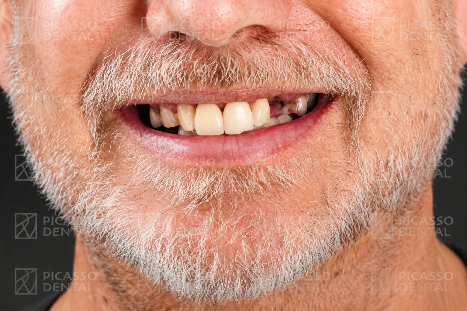 Before
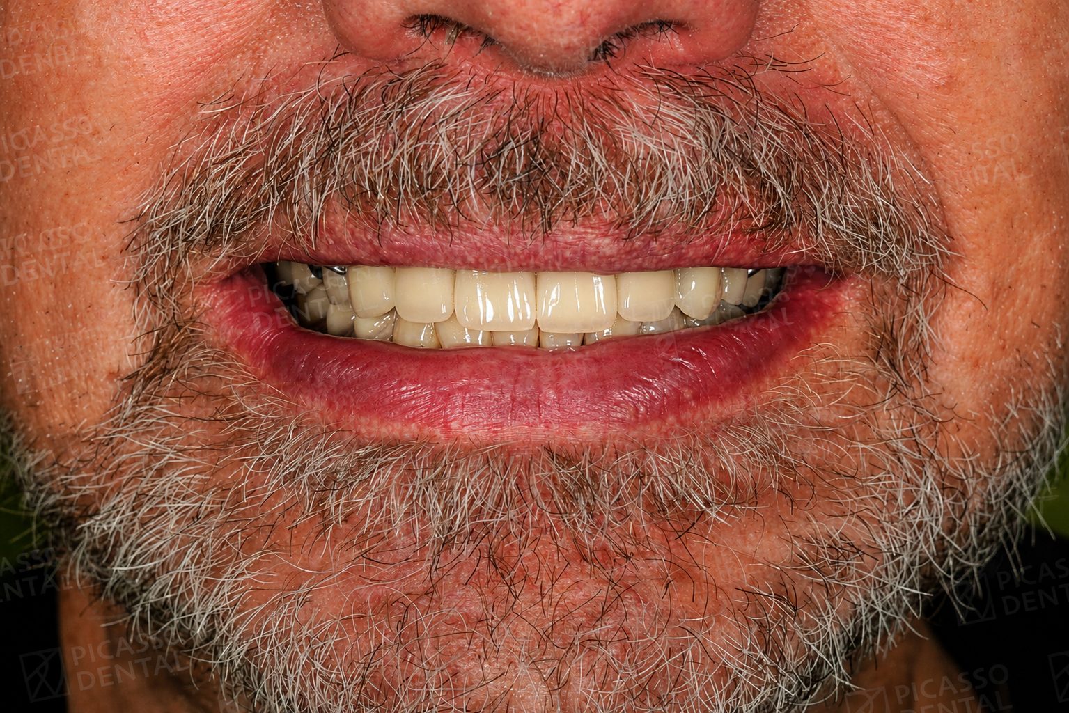 After
AfterCrown-protected root canal tooth. Biting force fully restored.
Frequently Asked Questions
Is the tooth dead after a root canal?
No. The pulp is removed, but the tooth itself is not dead. The tooth remains attached to the jaw by the periodontal ligament (PDL), which has its own independent blood and nerve supply. The PDL nourishes the root surface, holds the tooth in place, and gives you pressure-feedback when you bite. A root canal removes the internal pulp. It does not remove the external life support.
What does the periodontal ligament do after a root canal?
The periodontal ligament (PDL) is a thin band of fibrous tissue between the tooth root and the surrounding bone. It anchors the tooth, absorbs shock during chewing, supplies blood to the cementum on the outer root surface, and contains mechanoreceptors that detect bite pressure and position. After a root canal, the PDL continues all of these functions normally. The tooth is functionally alive at the bone interface even though the inner pulp is gone.
How long does a root canal-treated tooth last?
With a properly placed crown and routine maintenance, survival rates from large outcome studies are: 95 percent at 5 years, 85 to 90 percent at 10 years, 80 to 85 percent at 20 years, and 70 to 75 percent at 30 years. Many patients still have their root canal-treated teeth 40 or 50 years after the procedure. The key factors are quality initial treatment, a well-sealed crown, good oral hygiene, and 6-monthly review.
Can I still feel pressure on a root canal tooth?
Yes. You feel pressure normally because the periodontal ligament around the root is intact and the PDL contains the pressure-sensing nerves. What you lose is temperature sensitivity (hot and cold) because those nerves were inside the pulp, which has been removed. This is why root canal teeth need 6-monthly checkups: they cannot warn you about new cavities through temperature pain the way live teeth can.
Why does a root canal tooth sometimes darken?
A small percentage of root canal-treated teeth, especially front incisors, gradually darken to a grey or yellow shade over years. This happens because residual blood breakdown products inside the dentin tubules stain the tooth from within, and because internal fluid circulation from the pulp is absent. It is a cosmetic change, not infection or decay. Internal bleaching or a veneer or crown can restore the appearance.
Why do root canal teeth need a crown?
The pulp removal and the access cavity together leave the tooth structurally weaker than a vital tooth. Over time the dentin becomes more brittle without internal hydration. A crown holds the remaining structure together and distributes biting forces evenly. For molars and premolars, the published 5-year fracture rate without a crown is 15 to 30 percent. With a crown, it drops to 1 to 2 percent. See why a crown after root canal for the full reasoning.
What does a root canal tooth eventually fail from?
Long-term failures are almost always mechanical or hygienic, not biological. The three failure pathways are: vertical root fracture (especially in uncrowned posterior teeth), recurrent decay around the crown margin if oral hygiene is inadequate, and gum disease loosening the PDL attachment. A truly infection-driven biological failure is now rare with modern endodontic technique. Most root canal teeth do not die biologically; they wear out the way any restored tooth does.
Have a root canal tooth that needs a review?
A follow-up appointment at Picasso Dental includes X-ray assessment of the periapical area and PDL space, crown seal integrity check, and a frank conversation about whether the tooth is on a long-survival trajectory. No obligation, six branches across Hanoi, Da Nang, Ho Chi Minh City, and Da Lat.
Est. 2013 · Six branches · 70,000+ patients · 65+ countries