Dental Implants
Dental Implant Bone Loss:
Causes, Warning Signs, and Treatment
Some bone remodelling around a new implant is normal and expected. Progressive bone loss is not. Understanding the difference, recognising the early warning signs, and acting promptly determines whether a manageable situation becomes implant loss.
The short version
- 0.5 to 1 mm of crestal bone remodelling in the first year after implant placement is normal and expected.
- Bone loss exceeding 2 mm in the first year, or continuing to progress year on year, is pathological and needs intervention.
- Peri-implantitis (bacterial infection around the implant) is the primary cause of progressive bone loss.
- Early-stage peri-implantitis responds well to non-surgical cleaning and antimicrobial treatment if caught before significant bone is lost.
- Annual X-ray monitoring is the most reliable way to detect bone loss before it becomes visible or symptomatic.
Normal bone remodelling versus pathological bone loss
After an implant is placed, a small amount of crestal bone remodelling occurs in the first year as the soft tissue establishes a stable biological seal around the implant neck. This remodelling, typically 0.5 to 1 millimetre, is a normal biological response and does not indicate a problem.
Pathological bone loss, by contrast, is progressive. It exceeds 2 millimetres in the first year and continues beyond that threshold in subsequent years. This type of bone loss indicates peri-implantitis or another ongoing mechanical or biological problem that requires intervention. The distinction matters because early intervention is far more effective than late intervention.
Common causes of bone loss around implants
Peri-implantitis is the primary cause of progressive bone loss and the most common late implant complication. It is an inflammatory condition caused by bacterial biofilm accumulating around the implant, structurally similar to periodontitis around natural teeth. Risk factors include poor oral hygiene, a history of periodontal disease, smoking, and diabetes.
Occlusal overload occurs when excessive biting forces are applied to an implant, particularly during the osseointegration period. This disrupts bone formation and can cause the implant to loosen over time. Patients who grind their teeth are at particular risk. A custom nightguard protects against this and is strongly recommended for bruxers once the final crown is fitted.
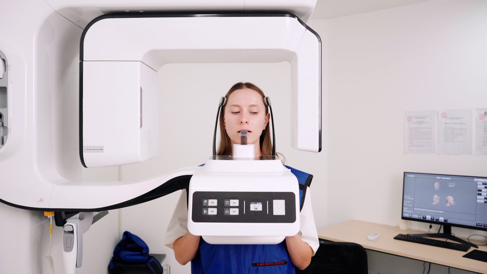
Implant position errors create biomechanical stress that promotes bone loss over years. An implant placed too close to an adjacent tooth, too shallow, or at an incorrect angle distributes chewing forces unevenly. This is why CBCT-guided surgical planning is not optional at Picasso Dental; it is the standard for every implant case.
Insufficient initial bone volume means the implant has inadequate bone surrounding it from the start. When a patient proceeds without adequate bone grafting, the thin margins of bone are more susceptible to resorption over time. A thorough pre-surgical CBCT identifies this issue before placement.
Systemic factors including poorly controlled diabetes, osteoporosis, certain medications (particularly bisphosphonates), and smoking all impair bone healing and increase susceptibility to bone loss around implants over time.
"During my total 4 to 5 hours of treatment on the 2 days of visit, I feel completely relaxed and do not feel any slightest pain during the treatment whosoever."
Wilfred Koh, HCMC Thao Dien, Google review
Warning signs to watch for
Bone loss around an implant is often silent in its early stages, which is why regular X-ray monitoring is essential. However, there are clinical signs that may indicate a developing problem.
- Gum redness, puffiness, or bleeding around the implantEarly indicators of peri-implant inflammation. Bleeding on gentle probing or when you brush the implant site is a consistent early warning sign.
- Pain or discomfort when chewingPain that was not present before, particularly on biting, deserves prompt evaluation. A stable implant should not hurt during normal function.
- Visible changes to the gum lineRecession revealing the implant surface or the junction between the abutment and crown indicates significant tissue loss and should be assessed immediately.
- Looseness or movement in the crownAny visible movement in the implant crown is a serious sign that bone loss may have progressed to the point where the implant is no longer firmly integrated.
Concerned about bone loss around your implant? Send your latest X-rays and describe your symptoms. We will review and advise, whether the implant was placed at Picasso or elsewhere.
Send X-rays for review →Diagnosis and treatment
Diagnosing peri-implantitis and quantifying bone loss requires clinical examination combined with X-ray assessment. We measure probing depths around the implant, assess bleeding on probing, and compare current X-rays with baseline images taken at implant placement. Cone beam CT scanning provides three-dimensional assessment of bone levels when the extent of involvement is unclear from two-dimensional films.
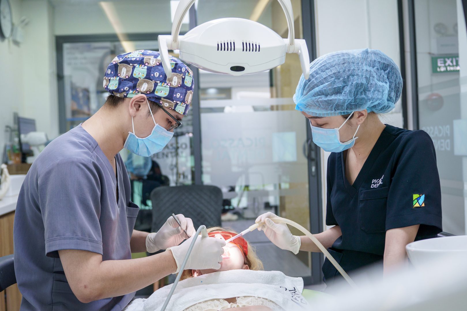
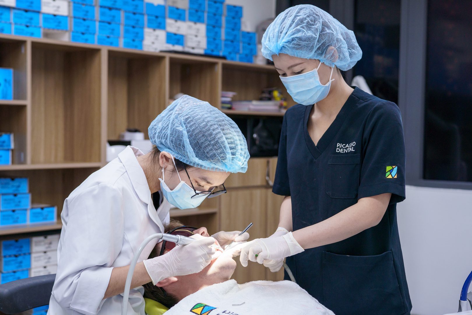
Non-surgical treatment is appropriate for early-stage peri-implantitis. Professional cleaning of the implant surface using specialised instruments removes the biofilm and calculus that drive the inflammatory process. Antimicrobial irrigation and, in some cases, local antibiotic delivery into the implant pocket complement mechanical cleaning. Rigorous home care instruction follows every treatment visit.
Surgical treatment is needed when non-surgical measures fail to resolve the infection or when bone loss is significant. Surgery allows direct access to the implant surface for thorough decontamination. Depending on the defect shape, bone grafting may be performed simultaneously to regenerate lost bone. Guided bone regeneration using barrier membranes can support new bone formation in contained defects.
Implant removal becomes necessary when bone loss is so extensive that the implant cannot be saved. After removal, the site heals, grafting is performed as needed, and re-implantation is planned once conditions are favourable. While not the outcome anyone wants, a failed implant can be successfully replaced in the majority of cases once the underlying cause is addressed.
Prevention is the best strategy
The most effective management of implant bone loss is preventing it from becoming significant. Meticulous daily oral hygiene around the implant, professional cleaning every six months, avoiding smoking, and managing systemic conditions keep the implant environment healthy. Annual X-ray monitoring allows early detection of even small amounts of bone loss before it becomes clinically significant. Patients who maintain regular appointments have vastly better long-term implant outcomes than those who attend only when they notice a problem.
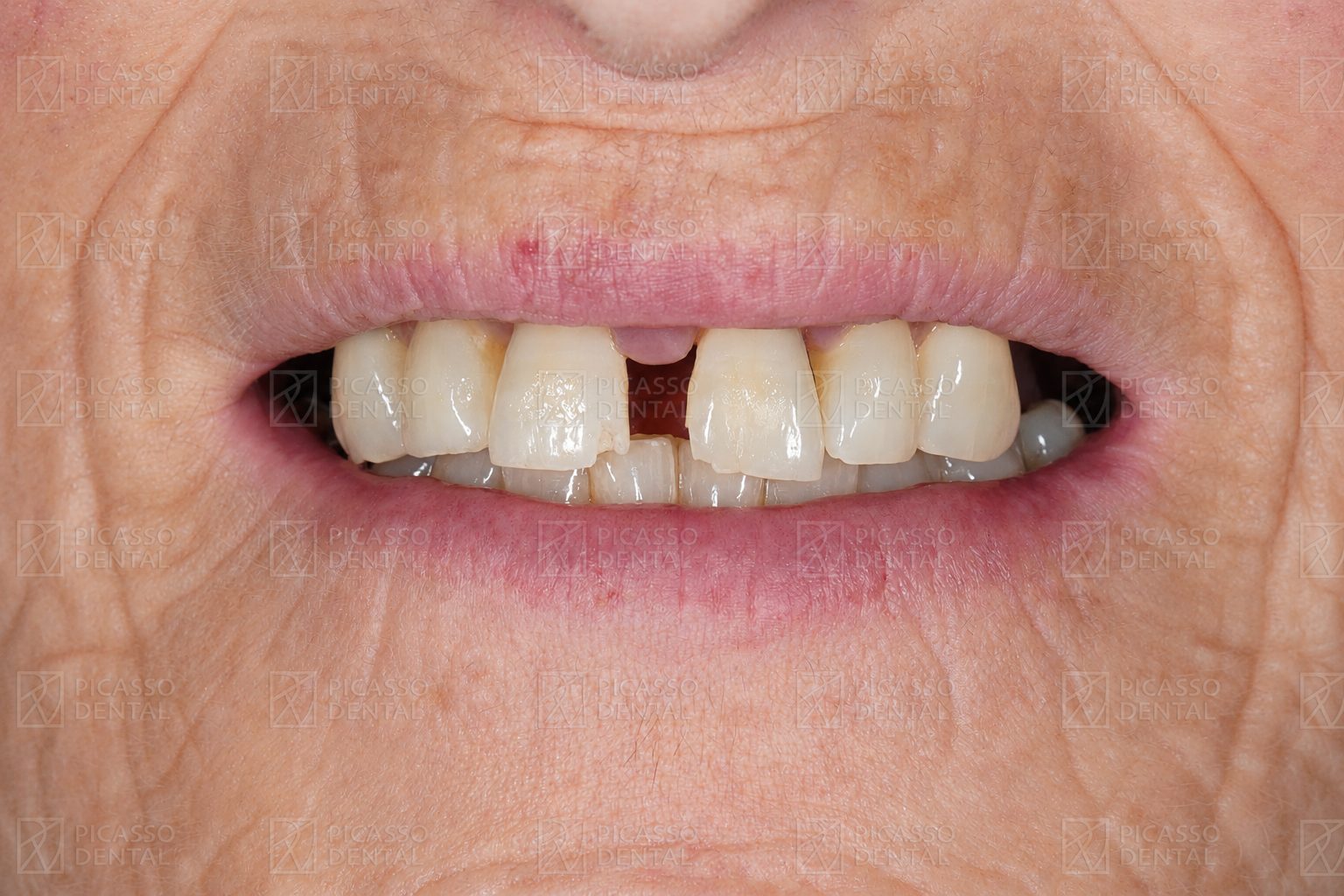 Before
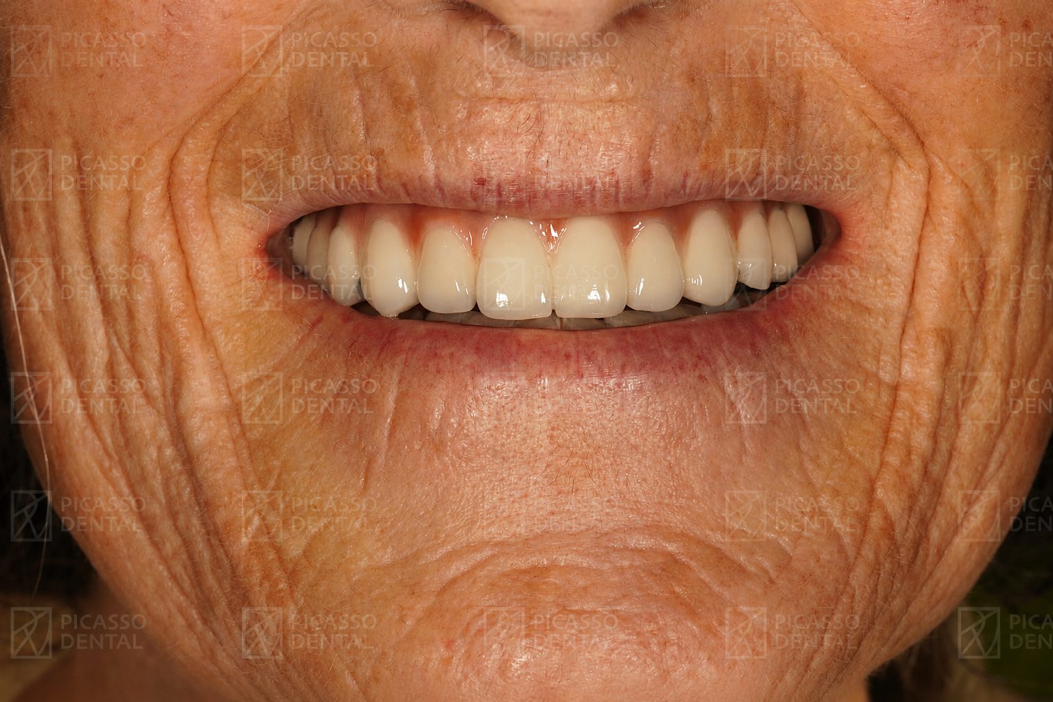 After
AfterFull rehabilitation, Hanoi Old Quarter
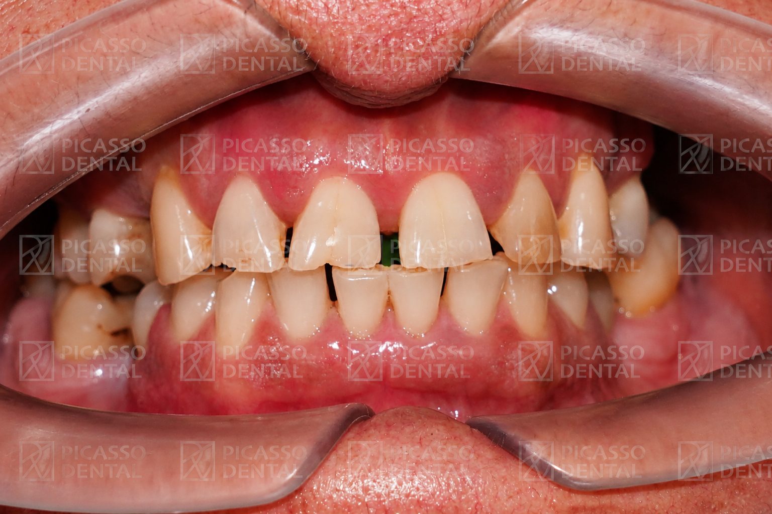 Before
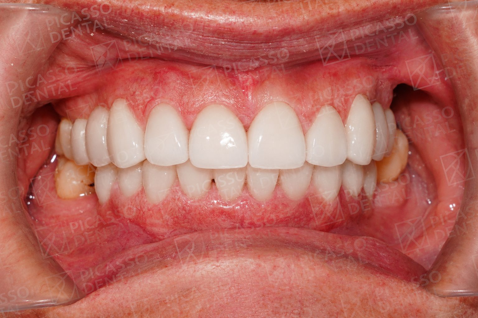 After
AfterPorcelain crowns, Da Nang
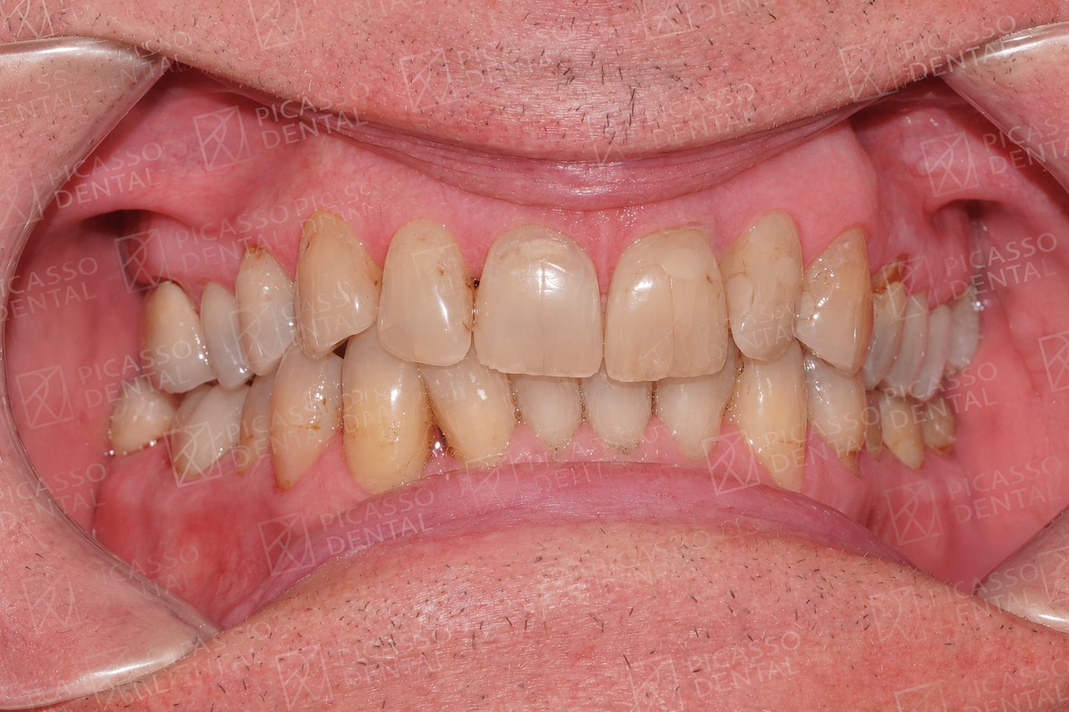 Before
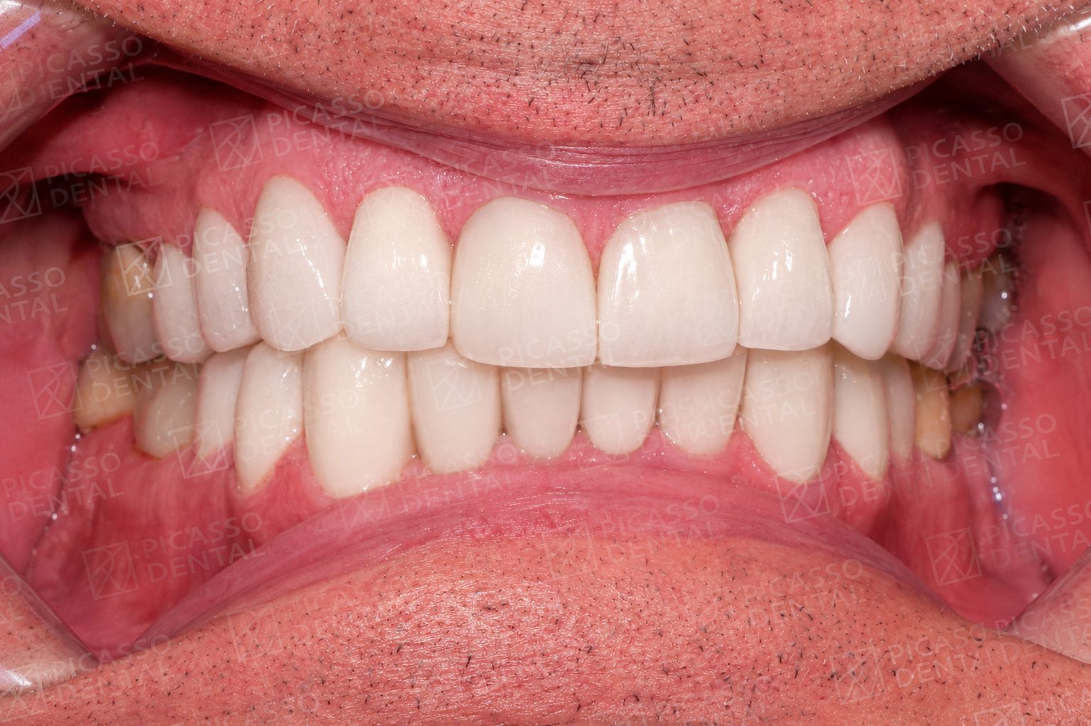 After
AfterCrown and implant, HCMC Thao Dien
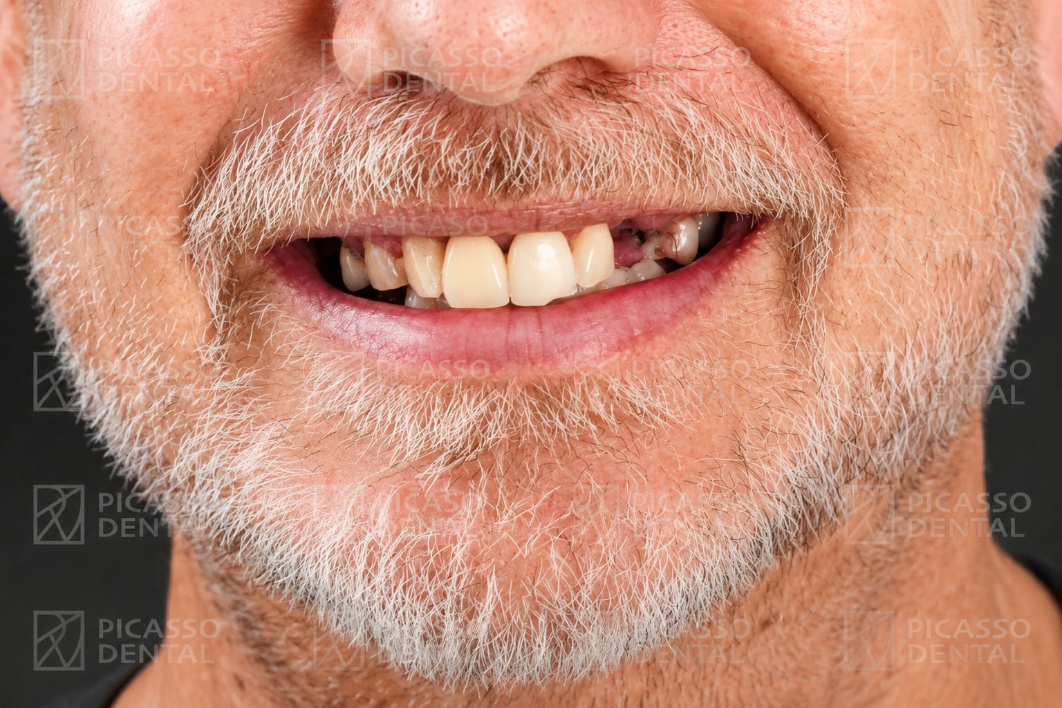 Before
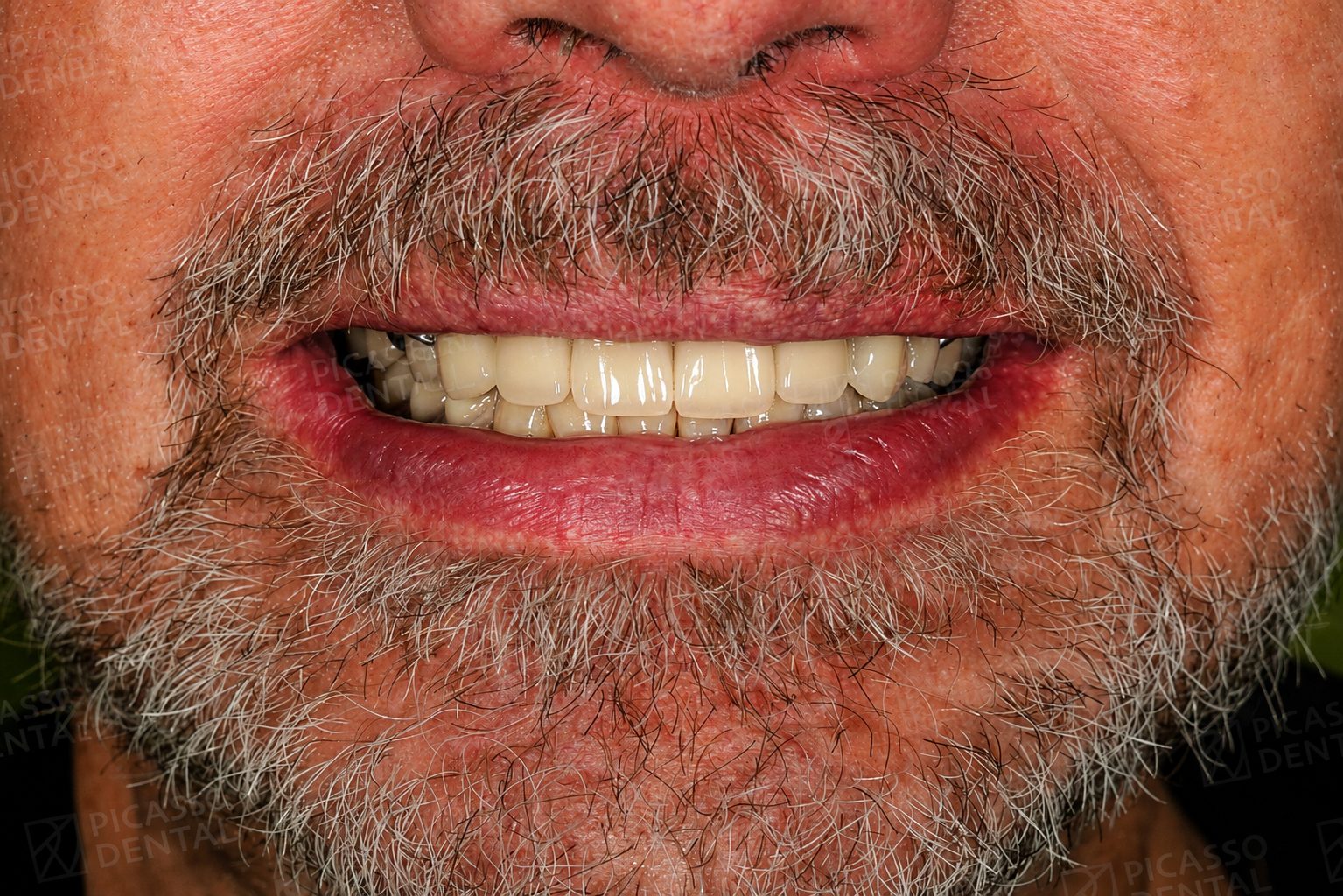 After
AfterCrown restoration, Hanoi Westlake
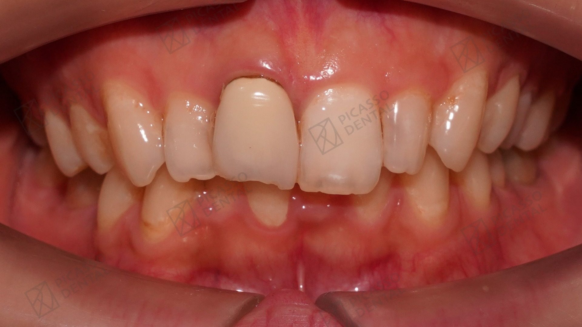 Before
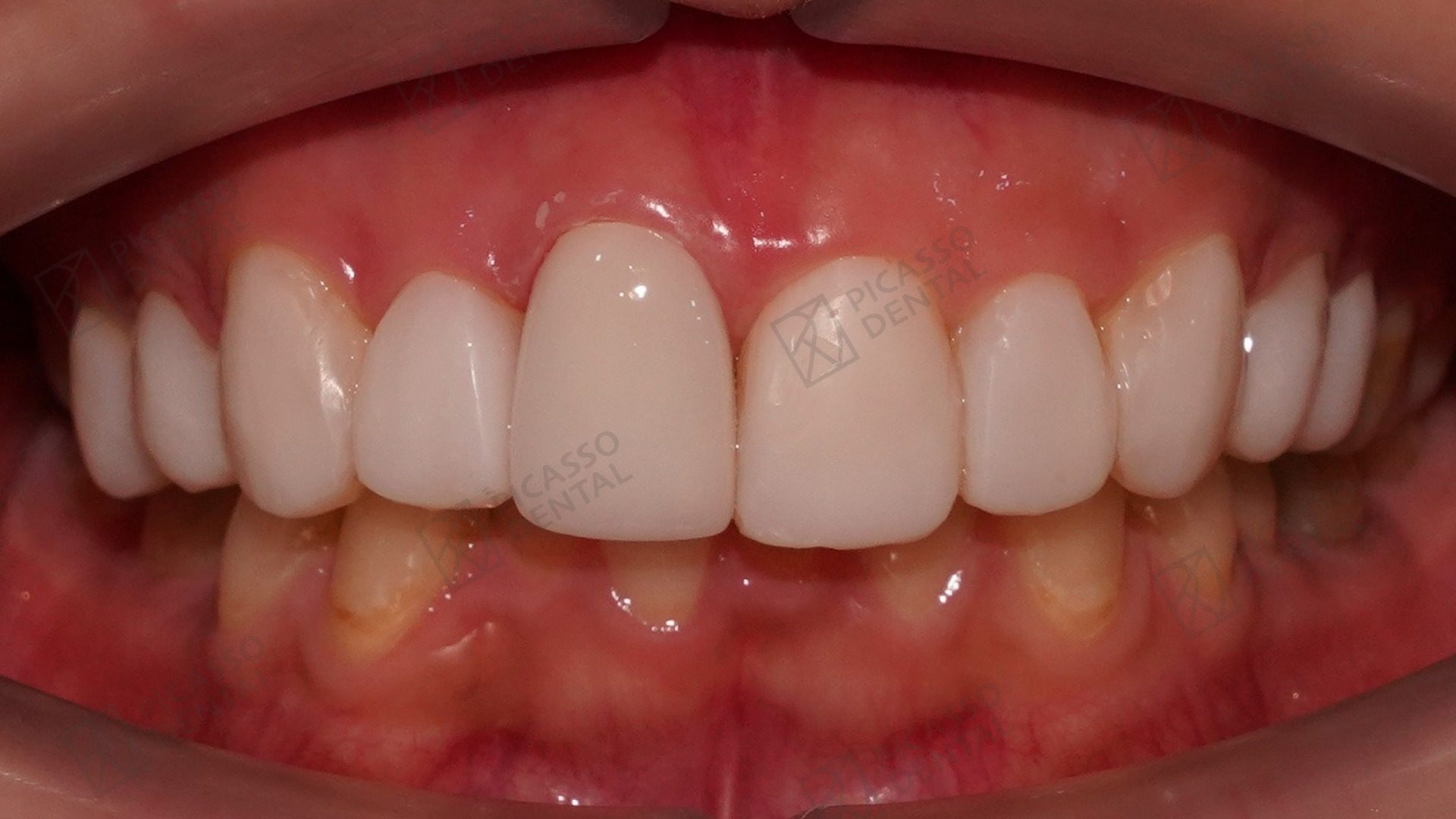 After
AfterSmile restoration, Da Nang Flagship
Frequently asked questions
How much bone loss around an implant is normal?
In the first year after implant placement, 0.5 to 1 millimetre of crestal bone remodelling is considered normal as the soft tissue establishes a stable seal around the implant. Bone loss exceeding 2 millimetres in the first year, or continuing to progress in subsequent years, is pathological and indicates a complication that needs intervention.
What causes bone loss around dental implants?
The primary cause is peri-implantitis, an inflammatory condition caused by bacterial biofilm accumulating around the implant. Other causes include occlusal overload from excessive biting forces or bruxism, implant position errors that create uneven force distribution, insufficient initial bone volume, and systemic factors such as uncontrolled diabetes, osteoporosis, smoking, and certain medications.
What are the warning signs of implant bone loss?
Early warning signs include gum redness, puffiness, or bleeding around the implant when you brush or probe. As bone loss progresses, you may notice pain or discomfort when chewing, visible changes to the gum line around the implant such as recession revealing the implant surface, and in advanced cases, looseness or movement in the implant crown.
How is implant bone loss diagnosed?
Diagnosis requires clinical examination combined with X-ray assessment. The dentist measures probing depths around the implant, assesses bleeding on probing, and compares current X-rays with baseline images taken at implant placement. Cone beam CT scanning provides three-dimensional assessment when the extent of bone loss is unclear from two-dimensional films.
Can implant bone loss be reversed?
Early-stage peri-implantitis can be halted and the surrounding environment stabilised with professional cleaning, antimicrobial treatment, and improved home care. In some cases, surgical intervention with guided bone regeneration can add new bone around the implant. Significant bone loss cannot be fully reversed, which is why annual X-ray monitoring and early intervention are essential.
What happens if an implant has too much bone loss to be saved?
When bone loss is so extensive that the implant cannot be maintained, removal is necessary. After removal the site heals, bone grafting is performed as needed, and re-implantation is planned once conditions are favourable. Most failed implants can be successfully replaced once the underlying cause is addressed.
How can I prevent bone loss around my implant?
Prevention requires meticulous daily oral hygiene around the implant (including flossing or water flosser use), professional cleaning every six months, avoiding smoking, managing systemic conditions such as diabetes, wearing a nightguard if you grind teeth, and attending annual X-ray check-ups so any early bone loss is detected before it becomes significant.
Start Here
Start with a brief.
Concerned about bone loss around an implant? Send your latest X-rays and describe your symptoms. The Picasso Dental implant team will review and advise on next steps. No obligation, 24-hour reply.