
General Dentistry
Can You Have Cavities
Between Teeth?
Yes, and They Are the Most Hidden
Interproximal cavities form where two teeth touch and are invisible without X-rays. Dr. Emily Nguyen explains why they are so common, how to detect them early, and the most effective prevention habits.

The short version
- Interproximal cavities form on the side surfaces of teeth where they contact each other, an area your toothbrush cannot reach.
- They are invisible to the naked eye and are detected only on bitewing X-rays during routine examinations.
- Food trapping, floss shredding, and cold sensitivity are the first symptoms, but early cavities often have no symptoms at all.
- Daily flossing with correct technique is the most effective single habit for preventing interproximal cavities.
- Small interproximal cavities require only a composite filling. Advanced ones may need an inlay, onlay, or root canal treatment if the nerve is involved.
Why the spaces between teeth are uniquely vulnerable
Your toothbrush reaches the outer faces of your teeth, the biting surfaces, and some of the inner surfaces facing your tongue or palate. What it cannot reach is the narrow contact zone where two adjacent teeth press against each other. This zone accounts for roughly 40 percent of the total tooth surface area, and it is the environment most favorable to cavity-forming bacteria.
When you eat, small food particles, particularly carbohydrates and sugars, become lodged in these contact areas. Saliva cannot easily flush these recessed spaces. Bacteria feed on the trapped food and produce organic acids as a byproduct. Those acids sit against the enamel for hours at a time, dissolving the mineral structure gradually. Without daily mechanical disruption of this bacterial film, the acid exposure accumulates over months into a cavity.

Back teeth are particularly at risk. Molars have tighter contacts than front teeth, do more chewing work that drives food between them, and are positioned further back in the mouth where flossing requires more effort and technique. This combination makes posterior interproximal cavities the most commonly treated type of decay in adult dentistry.
Why X-rays are essential for detection
Surface cavities on the chewing surfaces or outer faces of teeth can often be spotted visually during an examination. They discolor, feel sticky to an explorer, and may be visible as a dark spot. Interproximal cavities are different. They form on the side surfaces of teeth, completely hidden between two teeth. A cavity can progress from early enamel involvement to deep dentin involvement without ever being visible in the mouth.
Bitewing X-rays, the small films you bite down on during a dental check-up, are specifically designed to capture the contact areas between teeth in the posterior region. Early interproximal cavities appear as faint radiolucent (dark) areas on the X-ray within the enamel. Larger cavities show as clearly defined shadow areas penetrating the dentin. This X-ray evidence is often the only way to detect these cavities before symptoms appear.

Patients who attend regular examinations and have routine bitewing X-rays every 12 to 24 months catch interproximal cavities when they are small, still confined to the enamel or superficial dentin, and treatable with a simple filling. Patients who skip X-rays for years often present with cavities that have penetrated deeply, sometimes reaching the pulp chamber and requiring root canal treatment rather than a filling.
Warning signs that suggest a cavity between teeth
Early interproximal cavities are silent. The first detectable signs usually appear when the cavity has progressed through the enamel into the dentin layer beneath:
- Food consistently trapping between specific teeth A cavity creates a rough surface or small hole where smooth enamel used to be. Food wedges into this irregularity after every meal, in the same location each time. Many patients notice this before any pain develops and dismiss it as a problem with tooth spacing rather than recognizing it as a cavity symptom.
- Floss shredding or catching in the same spot Healthy enamel is smooth. A cavity in the contact area creates roughness or a defect that catches and tears floss. If your floss shreds consistently between the same two teeth, that is a sign that something is wrong, either a cavity, a rough filling edge, or calculus. All three need professional attention.
- Sensitivity to cold, sweet, or acidic foods Once decay reaches the dentin, the exposed dentinal tubules transmit temperature and chemical stimuli to the nerve. Sensitivity concentrated on one side of one specific tooth, rather than general mouth sensitivity, points to an interproximal cavity in that area. The sensation is typically a sharp twinge rather than a prolonged ache.
- Pain when biting down A more advanced cavity that has extended significantly into the dentin can cause pain when the teeth press together and compress the affected area. This symptom indicates a larger cavity that likely requires more than a simple filling.
"During my total 4 to 5 hours of treatment on the 2 days of visit, I feel completely relaxed and do not feel any slightest pain during the treatment whosoever."
Wilfred Koh, HCMC Thao Dien, Google review
Prevention: flossing technique matters more than frequency
Daily flossing is the non-negotiable foundation of interproximal cavity prevention. Toothbrush bristles simply cannot enter the contact space between teeth. Only floss provides the mechanical disruption of the bacterial film in that area. But the technique used is more important than simply doing it.
Most patients who floss use a snap-through technique: they pop the floss through the contact point, wiggle it once, and pull it back up. This cleans the contact point itself but misses the curved embrasure space below the contact, which is precisely where interproximal cavities begin. Correct technique requires curving the floss into a C-shape against each tooth, sliding it gently below the gumline, and moving it up and down several times before repositioning it against the adjacent tooth. Each side of the contact is treated separately.
Suspect a cavity between teeth? Describe your symptoms and a Picasso Dental clinician will respond within 24 hours with an honest assessment.
Contact us →Water flossers are a useful supplement, particularly for patients with braces, bridges, or very tight tooth contacts where string floss is difficult to use. The pressurized water stream removes food debris and reduces bacterial counts. They are not, however, a complete substitute for string floss. The friction of the string against the tooth surface is more effective at disrupting the bacterial biofilm than water pressure alone. Using both is the most thorough approach.

Treatment options by cavity size
The treatment required for an interproximal cavity depends directly on how far the decay has progressed when it is detected. This is the clearest argument for regular X-rays: small cavities require small interventions.
Early enamel cavities that have not yet penetrated into the dentin can sometimes be remineralized using fluoride treatments and improved home care, avoiding any drilling. Once decay has entered the dentin, removal is required.
Small to medium interproximal cavities are treated with composite resin fillings. After removing all infected tooth structure, the cavity is cleaned, conditioned, and filled with tooth-colored composite that bonds to the remaining enamel and dentin. Access requires careful technique because the space is narrow, but the procedure is straightforward and completed in a single appointment.
Large cavities that have destroyed significant dentin may need a ceramic inlay or onlay rather than a direct filling. These restorations are fabricated by a laboratory from impressions and bonded into place at a second appointment. They are stronger and more durable than large composite fillings under heavy chewing loads.
Cavities that have reached the dental pulp require root canal treatment before the tooth can be restored. The infected nerve tissue is removed, the canals cleaned and sealed, and the tooth restored with a filling or crown. A cavity that could have been treated with a 30-minute filling appointment becomes a multi-appointment case when left until nerve involvement occurs. The cost difference is significant, and the experience is more involved for the patient.
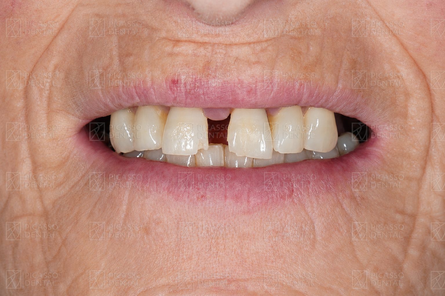 Before
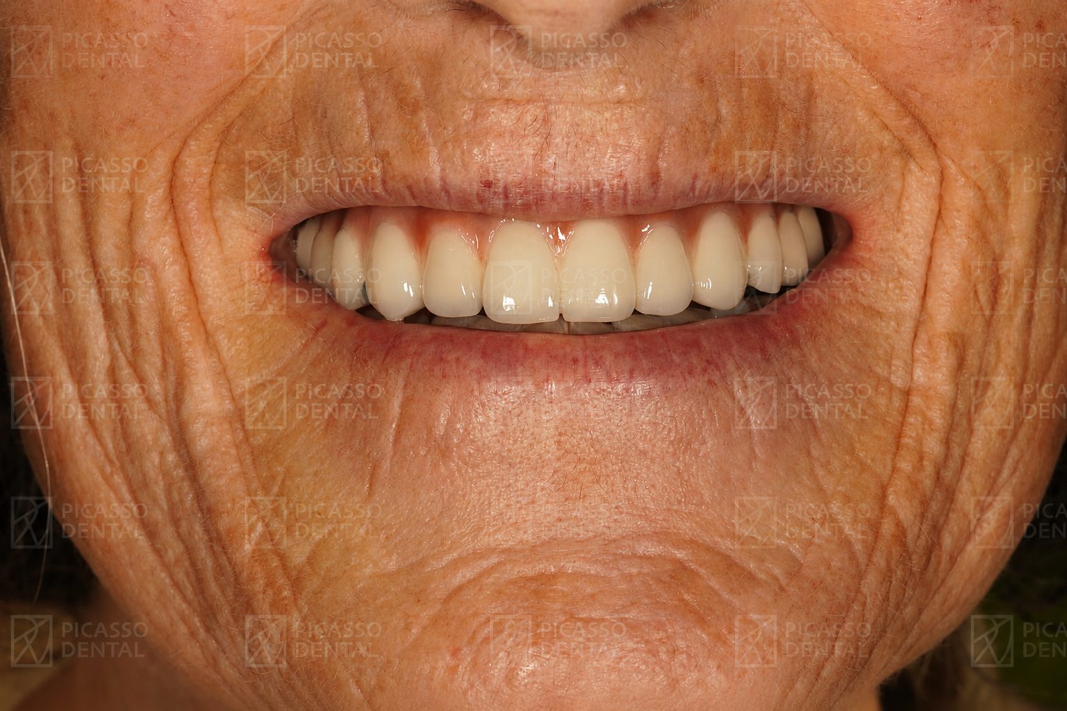 After
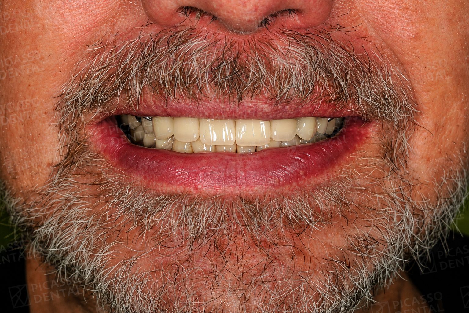
AfterFull rehabilitation, Hanoi Old Quarter
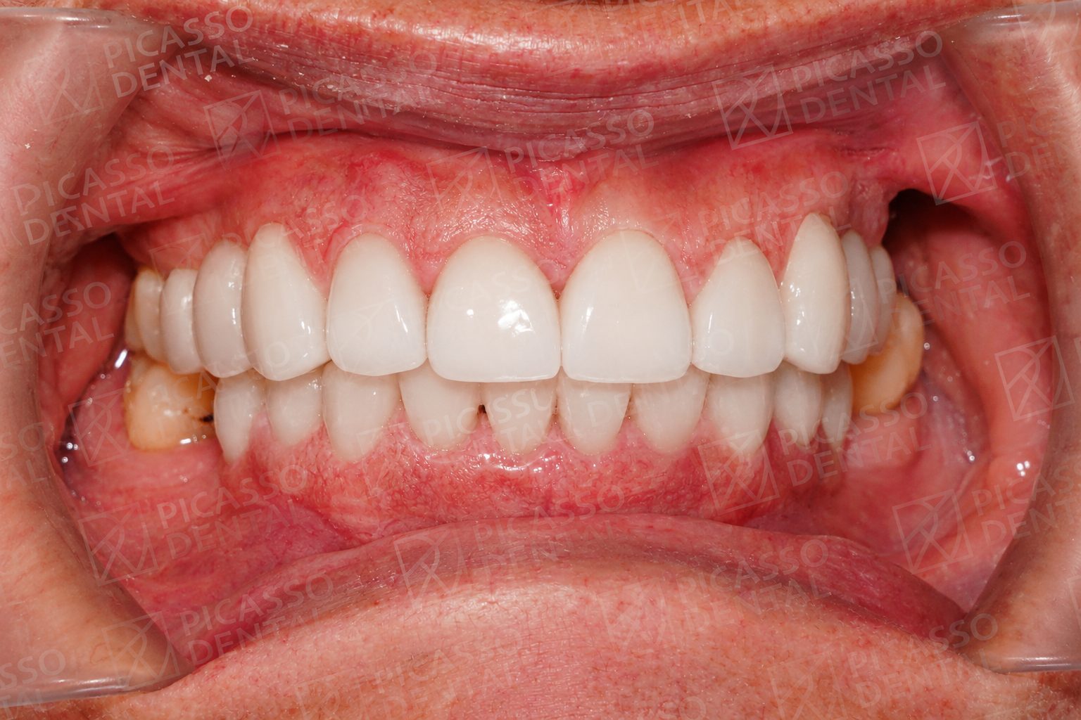
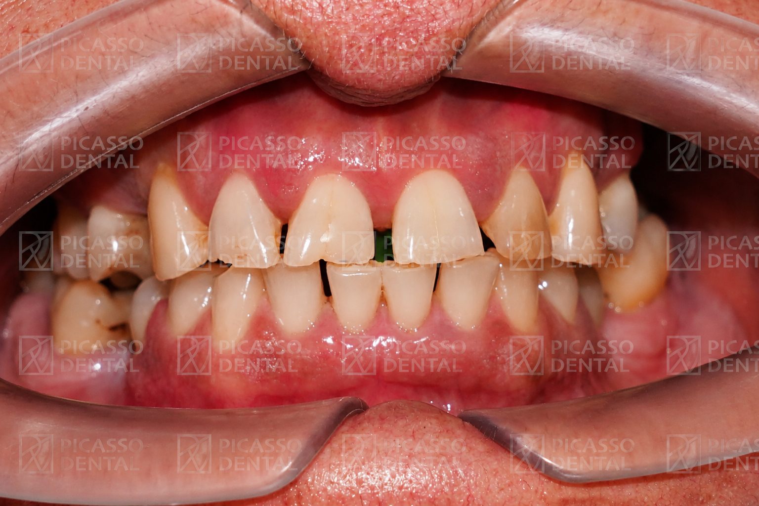 Before
 After
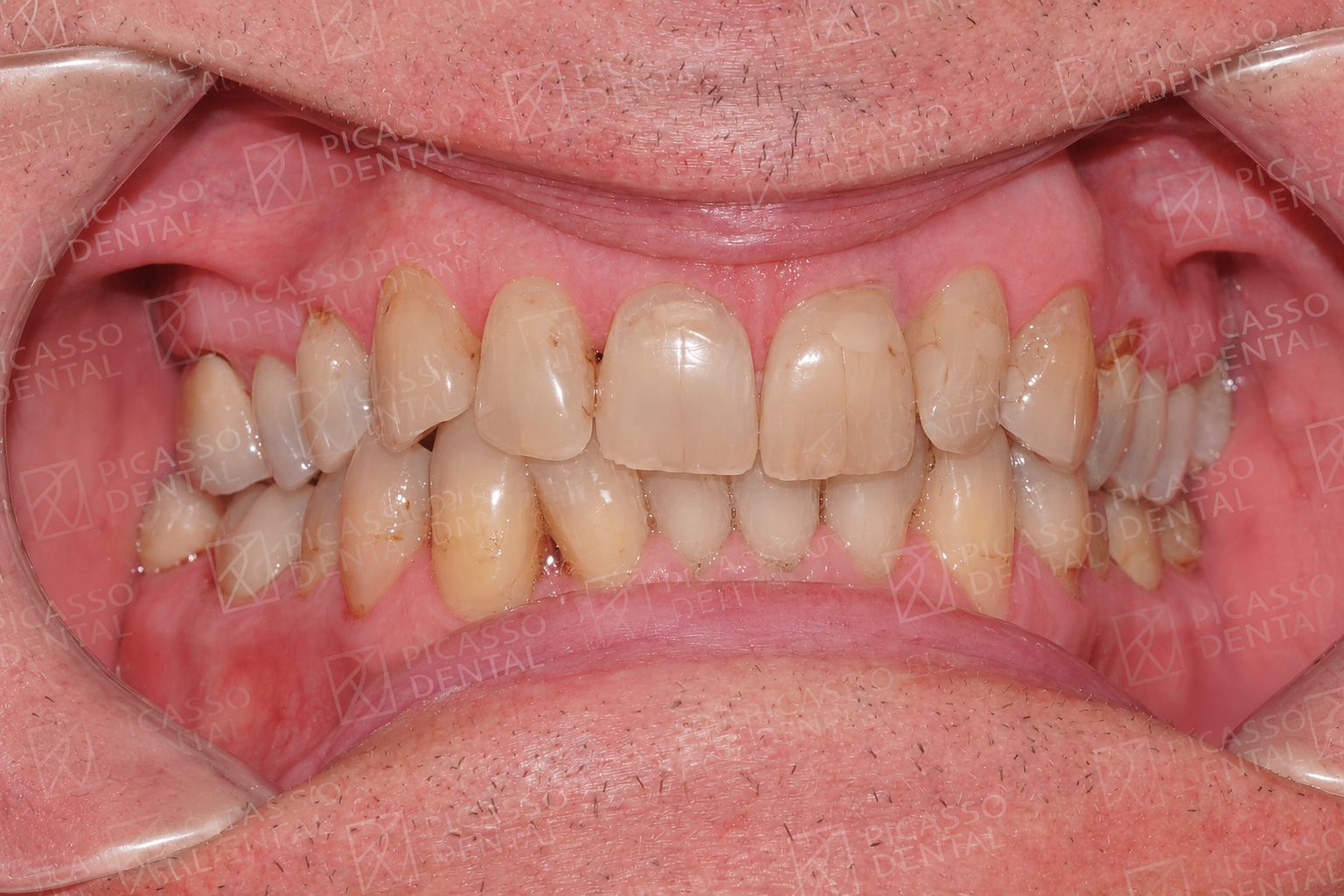
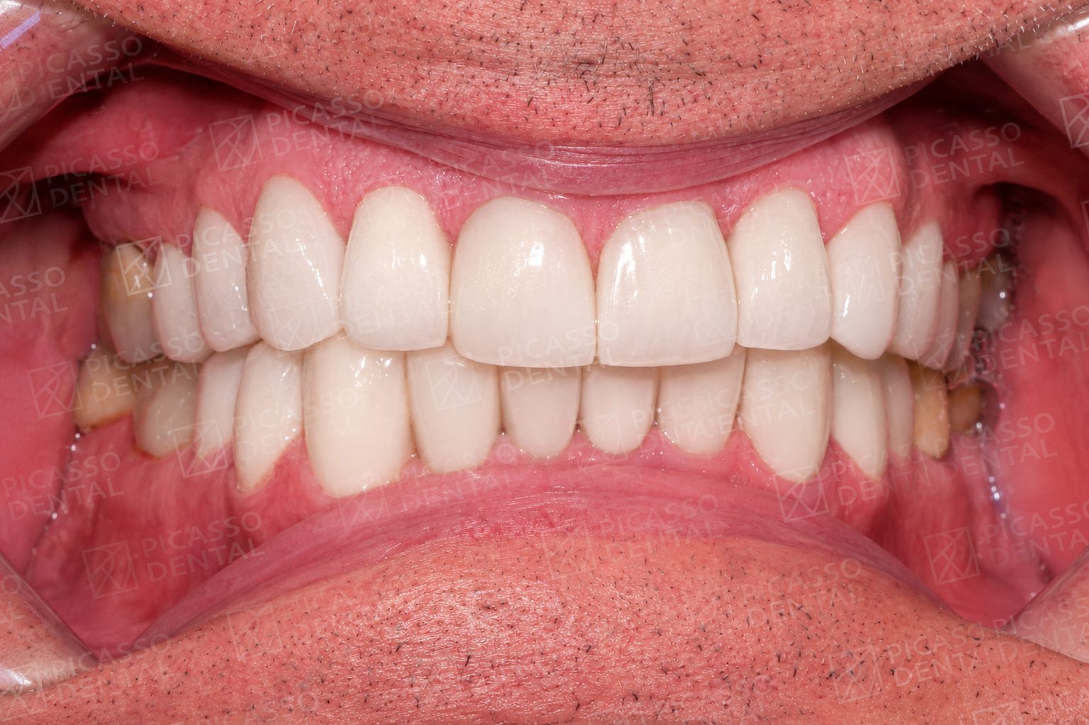
AfterPorcelain crowns, Da Nang
 Before
 After
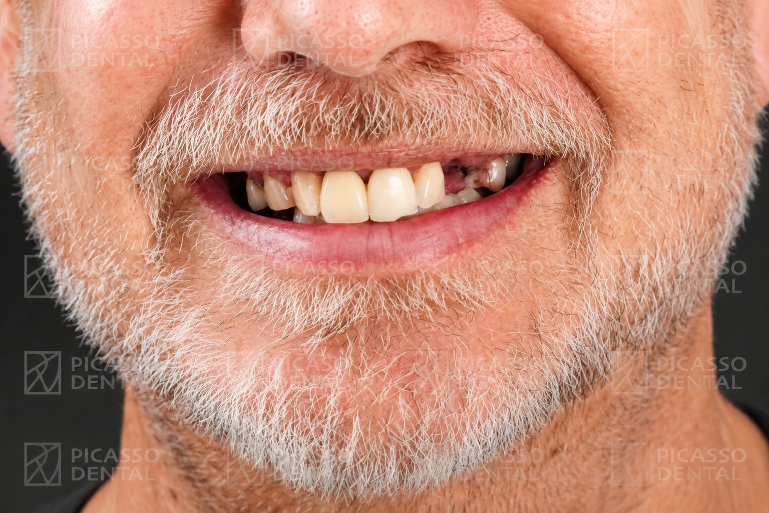
AfterCrown and implant, HCMC Thao Dien
 Before
 After
AfterCrown restoration, Hanoi Westlake
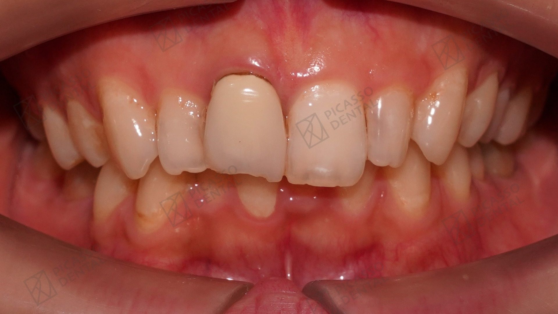 Before
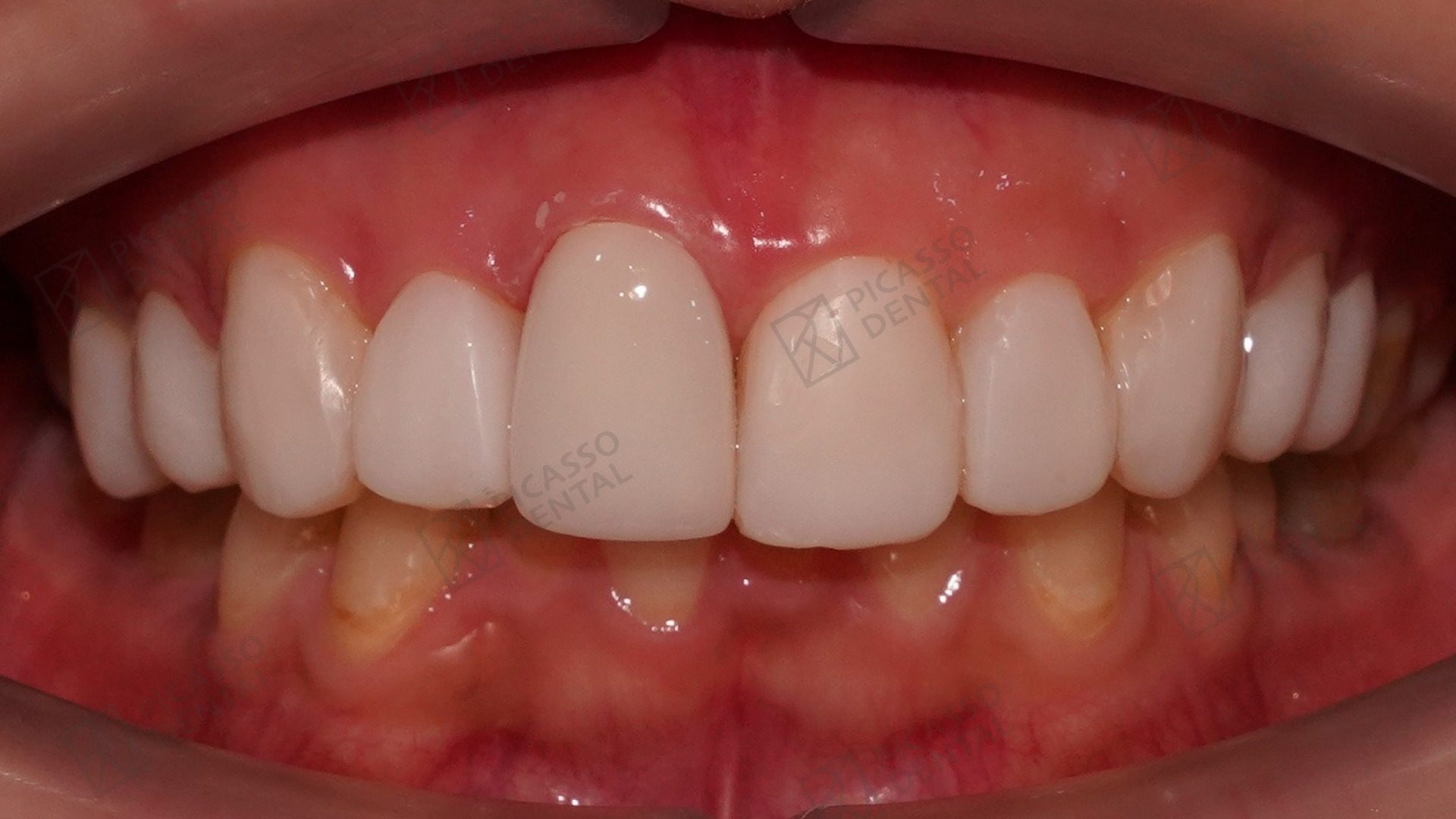 After
AfterSmile restoration, Da Nang Flagship
Frequently asked questions
Can you have cavities between teeth?
Yes. Interproximal cavities, which form on the side surfaces where two teeth contact each other, are among the most common types of decay. They are invisible to the naked eye and to toothbrush bristles, which is why they often grow undetected until an X-ray reveals them or symptoms appear.
How are cavities between teeth detected?
Bitewing X-rays are the primary tool for detecting interproximal cavities. These small X-ray films, taken during routine examinations, show the spaces between teeth where decay cannot be seen directly. Early interproximal cavities show as faint shadow areas on the X-ray. Advanced cavities that have broken through to a visible surface can sometimes be seen clinically, but the majority of early interproximal decay is found only on X-ray.
What are the symptoms of a cavity between teeth?
Early interproximal cavities have no symptoms. As they progress, the first signs include food consistently becoming trapped between specific teeth, floss shredding or catching in the same location, and sensitivity to cold, sweet, or acidic foods. Pain when biting down indicates a more advanced cavity that has reached the dentin. Very advanced cavities may cause spontaneous toothache as the decay approaches the nerve.
Can flossing prevent cavities between teeth?
Daily flossing is the most effective single habit for preventing interproximal cavities. Toothbrush bristles cannot reach the contact areas between teeth. Floss removes food particles and disrupts the bacterial film that produces acid. Correct flossing technique, curving the floss against each tooth in a C-shape and moving it up and down rather than just snapping it through the contact, is more important than the frequency of flossing.
How are cavities between teeth treated?
Small interproximal cavities are treated with composite resin fillings placed after removal of the decay. Access to the cavity requires careful technique because the space between teeth is narrow. Larger cavities may need ceramic inlays or onlays for greater durability. Cavities that have reached the nerve require root canal treatment before the tooth can be restored with a filling or crown.
How often should I get dental X-rays to check for cavities between teeth?
For adults with a normal cavity risk, bitewing X-rays every 12 to 24 months allow detection of interproximal cavities while they are still small and treatable with a simple filling. Patients with a higher cavity risk, including those with dry mouth, frequent sugar intake, or a history of many cavities, benefit from X-rays every 12 months. Children and teenagers with tight tooth contacts are typically X-rayed annually.
Can a water flosser prevent cavities between teeth?
Water flossers reduce food debris and bacterial counts between teeth and are particularly useful for patients with braces, bridges, or tight contacts where traditional floss is difficult to use. However, they are not a complete replacement for string floss. The mechanical disruption of the bacterial film against the tooth surface is better achieved with the friction of floss than with water pressure alone. Using both is the most effective approach.
Start Here
Start with a brief.
If you are experiencing food trapping, floss shredding, or tooth sensitivity, describe your symptoms and a Picasso dentist will respond within 24 hours. No obligation.