
Root Canal Treatment
Calcified Root Canal Treatment
at Picasso Dental:
The tools, the technique, and the honest odds
Calcified canals are among the most technically demanding procedures in endodontics. Here is exactly how they are treated, what equipment is essential, and what you should expect in terms of success rates.

The short version
- Calcified canals occur when mineral deposits narrow or block the hollow space inside the root, making standard root canal instruments unable to enter.
- A dental microscope at 8 to 20 times magnification is essential for locating and treating calcified canals safely.
- Ultrasonic instruments and EDTA chelating agent remove calcification without cutting tooth structure.
- Success rates are 70 to 90 percent, lower than standard root canals, depending on calcification severity and operator skill.
- When treatment is not possible, apicoectomy or extraction with implant replacement are the remaining options.
What calcification does to a root canal
Root canal calcification occurs when mineral deposits gradually narrow or completely block the hollow spaces inside tooth roots where nerves and blood vessels reside. This happens naturally with aging, as a protective response to trauma or prolonged grinding, after years of untreated decay approaching the nerve, or as a result of certain medical conditions. The canal space that should be hollow becomes partially or completely filled with hard calcified material.
What appears on X-rays as a tooth with no visible canal space actually does have canals present. They are simply blocked by calcification that obliterates their image on conventional radiographs. Young patients rarely have significant calcification. Older patients, particularly those over 50, commonly show some degree of calcification in teeth requiring root canal treatment. Teeth with a history of trauma, even minor impacts decades earlier, are particularly prone.
Calcification makes root canal treatment dramatically more difficult because I cannot simply clean out canals as with normal treatment. The calcified material must be carefully removed without weakening the tooth or perforating through the thin root wall into surrounding bone. This requires fundamentally different tools and techniques to standard endodontic treatment.
Why the dental microscope is non-negotiable
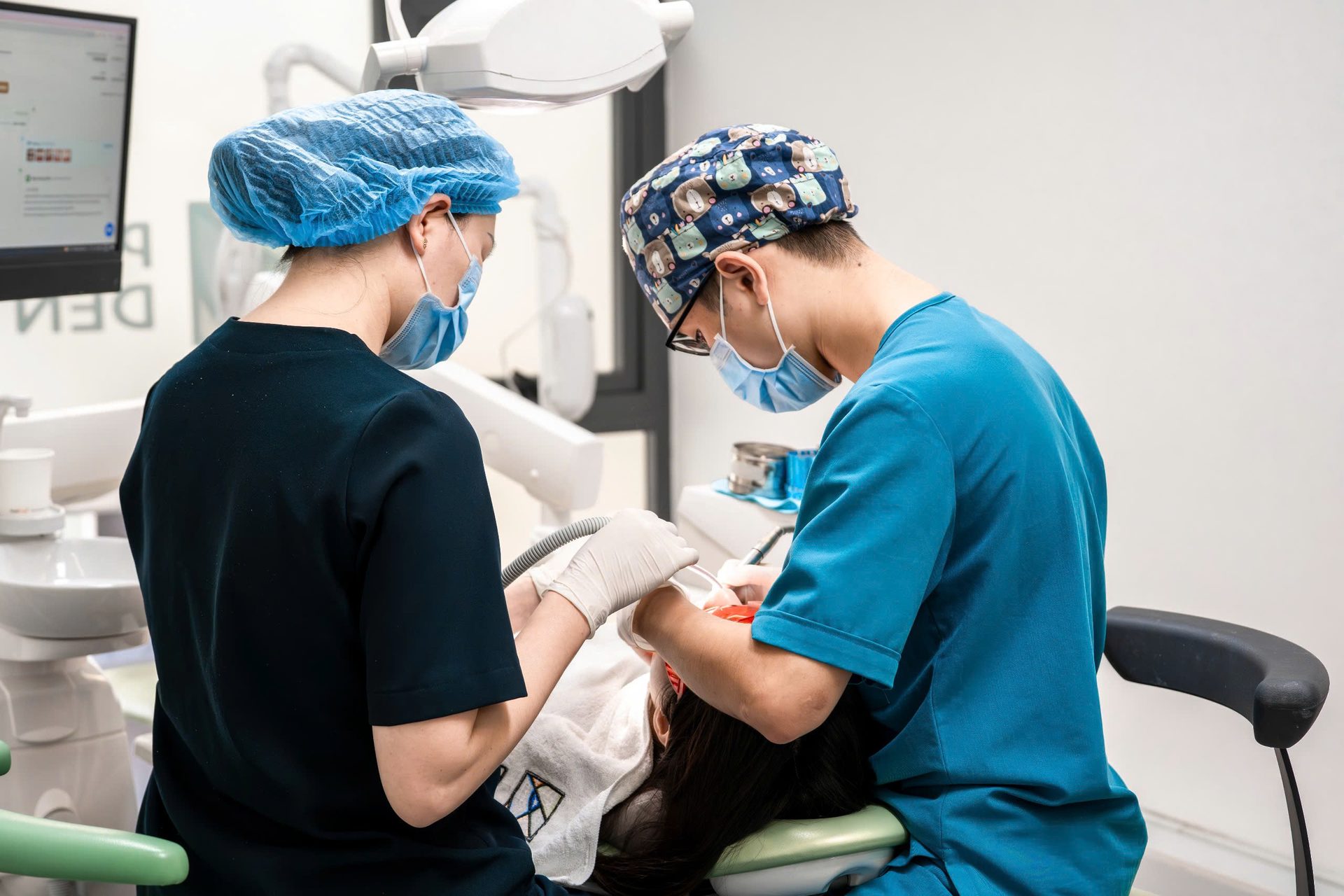
Treating calcified canals without a dental microscope is the most common reason these procedures fail at clinics that attempt them. The microscope provides 8 to 20 times magnification with intense co-axial illumination, allowing the clinician to see subtle colour changes and structural features inside the tooth that are completely invisible to the naked eye or through standard loupes.
Under microscope magnification, the clinician can identify the tiny opening where a calcified canal begins, a yellowish or brownish dot barely 0.1 millimetres in diameter, distinguish calcified tissue from surrounding dentine, and navigate safely without perforating the thin root wall. Without this level of detail, the dentist is navigating largely blind. Perforation and failure rates in calcified canal treatment without magnification are significantly higher.
Ultrasonic instruments and EDTA: the tools that make it possible
Ultrasonic tips vibrate at extremely high frequencies, typically between 25,000 and 30,000 cycles per second. This vibration breaks apart calcified material through mechanical energy without cutting the surrounding tooth structure. Different tip shapes are designed to access different canal configurations and depths. The precision of ultrasonic removal, compared to mechanical cutting instruments, reduces the risk of accidentally removing excess tooth structure and weakening the root.
EDTA, a chelating agent applied as a liquid or gel, chemically softens calcified material by binding the calcium ions within it. Applied for several minutes before mechanical instrumentation, EDTA makes the calcification more friable and easier to remove. It does not affect the healthy dentine surrounding the calcified material. The combination of ultrasonic mechanical action and EDTA chemical action produces better outcomes than either approach alone.
Sodium hypochlorite, the primary irrigating solution throughout root canal treatment, provides additional benefit in calcified cases by dissolving organic material trapped within the calcification matrix and eliminating bacteria introduced during the instrumentation process.
"I had four crowns, two cavities, and a root canal and it was as gentle and pain free as it could possibly be. More so than any dentist back in the states in my sixty years of seeing the dentist."
Kevin McCarthy, Hanoi Old Quarter
Been told your root canal is calcified or untreatable? A second opinion at Picasso Dental Clinic includes CBCT assessment and a clear success probability estimate before any commitment to treatment.
Book a second opinionTreatment timeline and what to expect
Calcified canal treatment typically requires two to three appointments. The first appointment is the most demanding: locating the canal opening, beginning calcification removal with ultrasonic instruments and EDTA, shaping as far as safely possible, irrigating thoroughly, and placing intracanal medication. This appointment takes two to three hours for a molar with multiple calcified canals. Extending the appointment adequately rather than rushing is essential to avoid the errors that cause perforations and treatment failure.
Between appointments, intracanal medication helps dissolve residual organic material within the remaining calcification and reduces bacterial colonisation. The second appointment, typically 60 to 90 minutes, completes canal cleaning and shaping and places the permanent obturation material. In complex cases with severely calcified anatomy, a third appointment may be necessary.
After treatment, a crown is placed over the treated tooth to protect it from fracture. Calcified teeth that have been treated successfully function and feel like any other root-canal-treated tooth. They do not require special care beyond the usual oral hygiene and six-monthly examinations.

When treatment is not the right choice
Not all calcified canals can be treated successfully, and attempting treatment on cases with very poor prognosis wastes the patient's time and money. Complete obliteration of all canals with no identifiable anatomy, anatomy that makes safe access impossible without near-certain perforation, or previous failed attempts that have already compromised the root structure are all situations where the honest recommendation is extraction and replacement.
Apicoectomy, surgical access to the root tip from outside the tooth, sometimes provides an alternative when the upper portion of the canal is accessible but the calcified tip portion cannot be reached from above. The calcified tip is removed surgically and the accessible portion of the canal is sealed. This surgical approach works in select cases and is worth discussing with the treating dentist when the conventional approach has limited prognosis.
Setting realistic expectations is part of responsible treatment planning. Success rates of 70 to 90 percent are good but not the 90 to 95 percent of non-calcified canals. Some patients proceed with treatment accepting the risk. Others, particularly when the tooth in question has limited remaining strategic value and replacement with an implant would be straightforward, choose extraction as the more predictable path. Both decisions can be correct depending on the specific case.
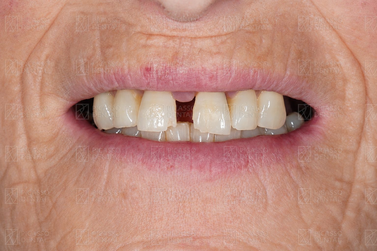 Before
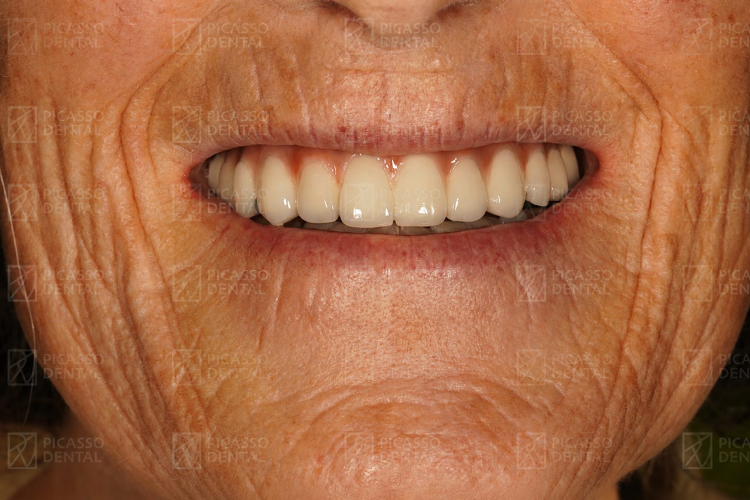 After
AfterFull rehabilitation, Hanoi Old Quarter
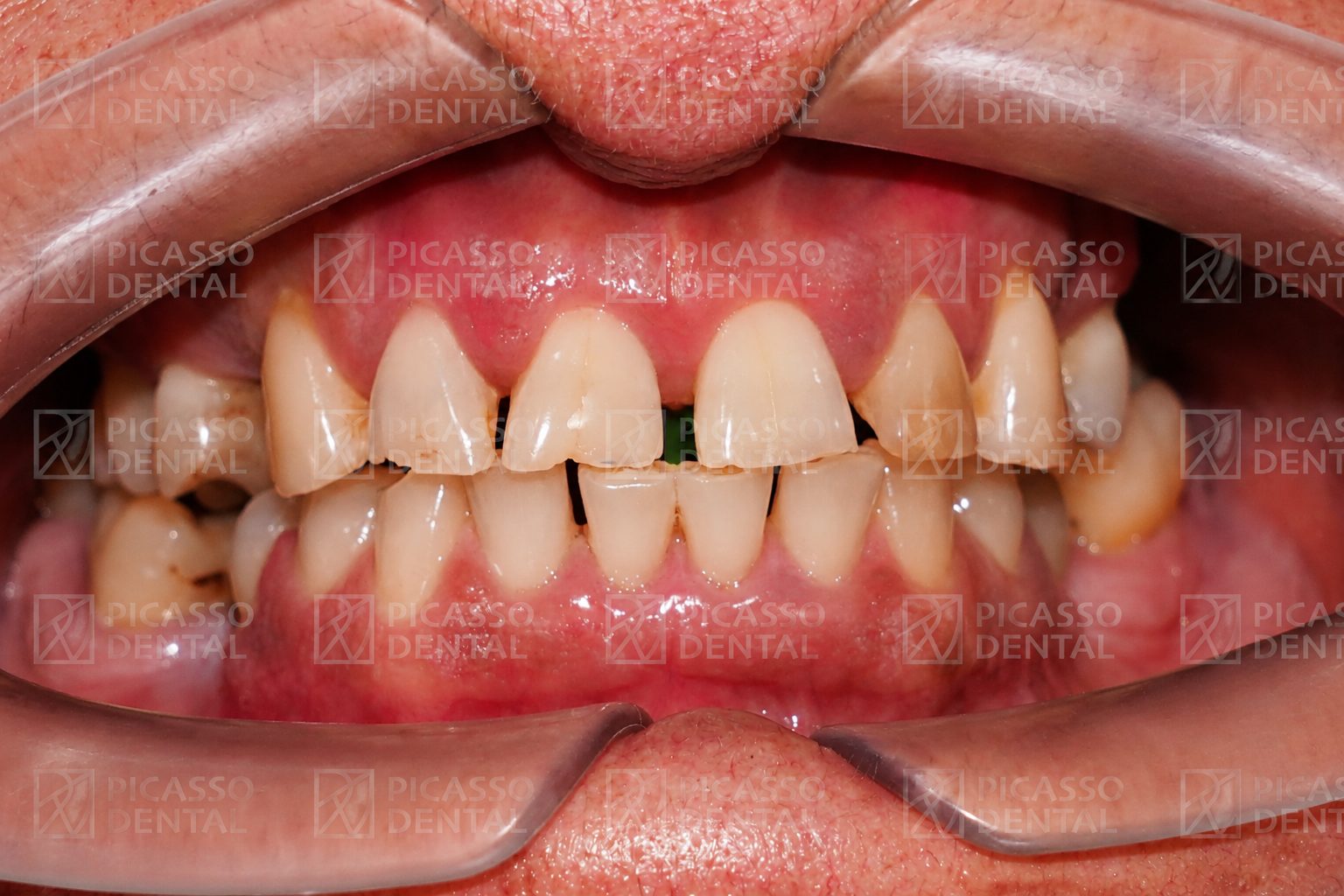 Before
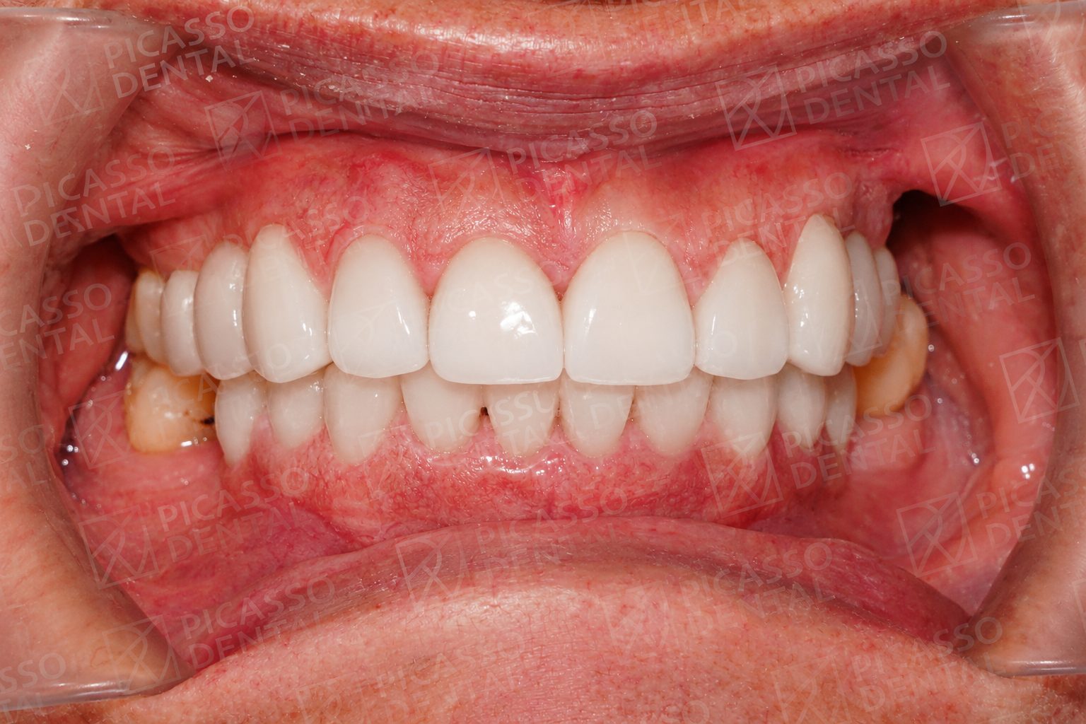 After
AfterPorcelain crowns, Da Nang
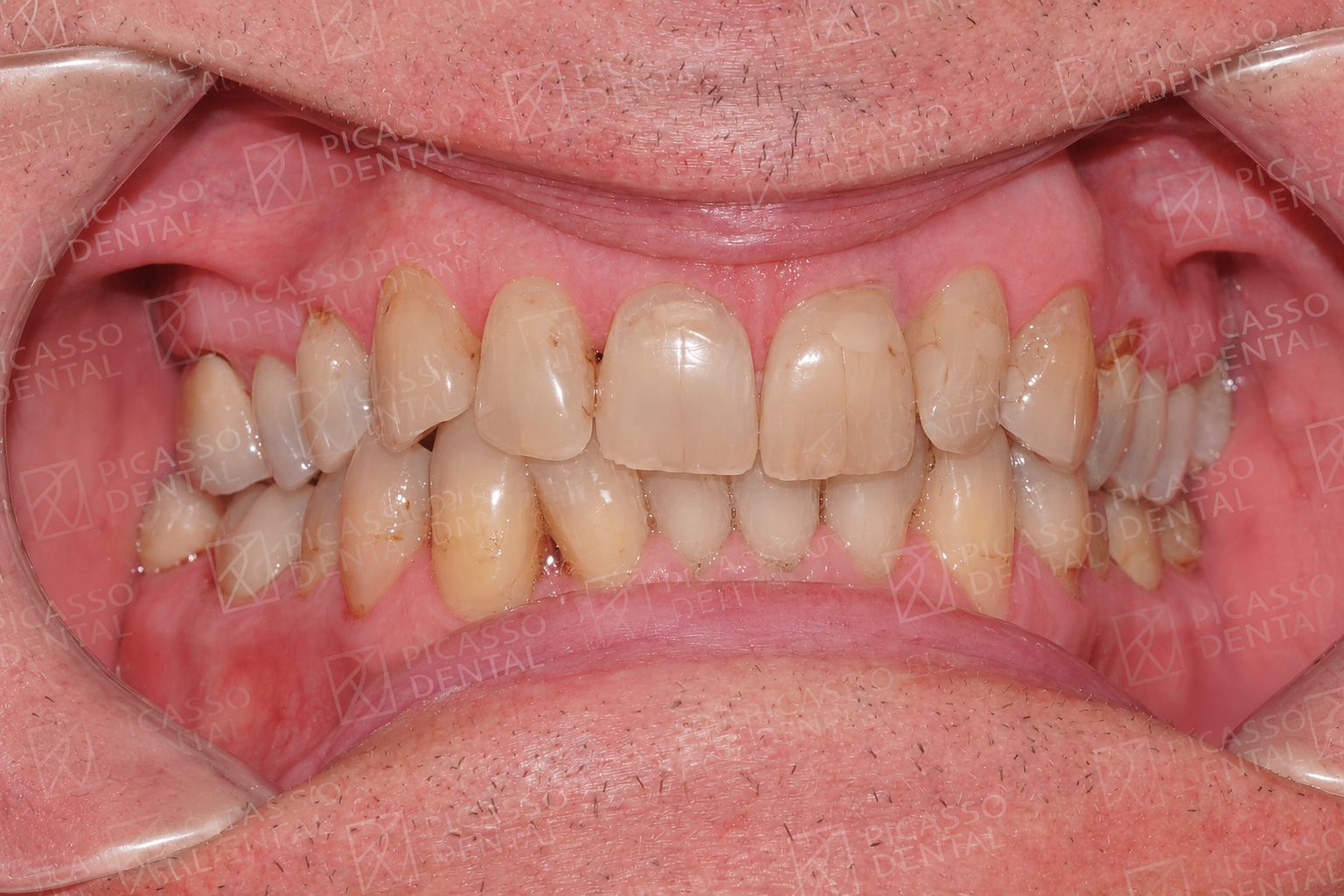 Before
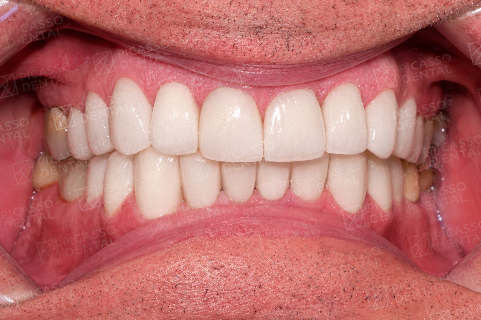 After
AfterCrown and implant, HCMC Thao Dien
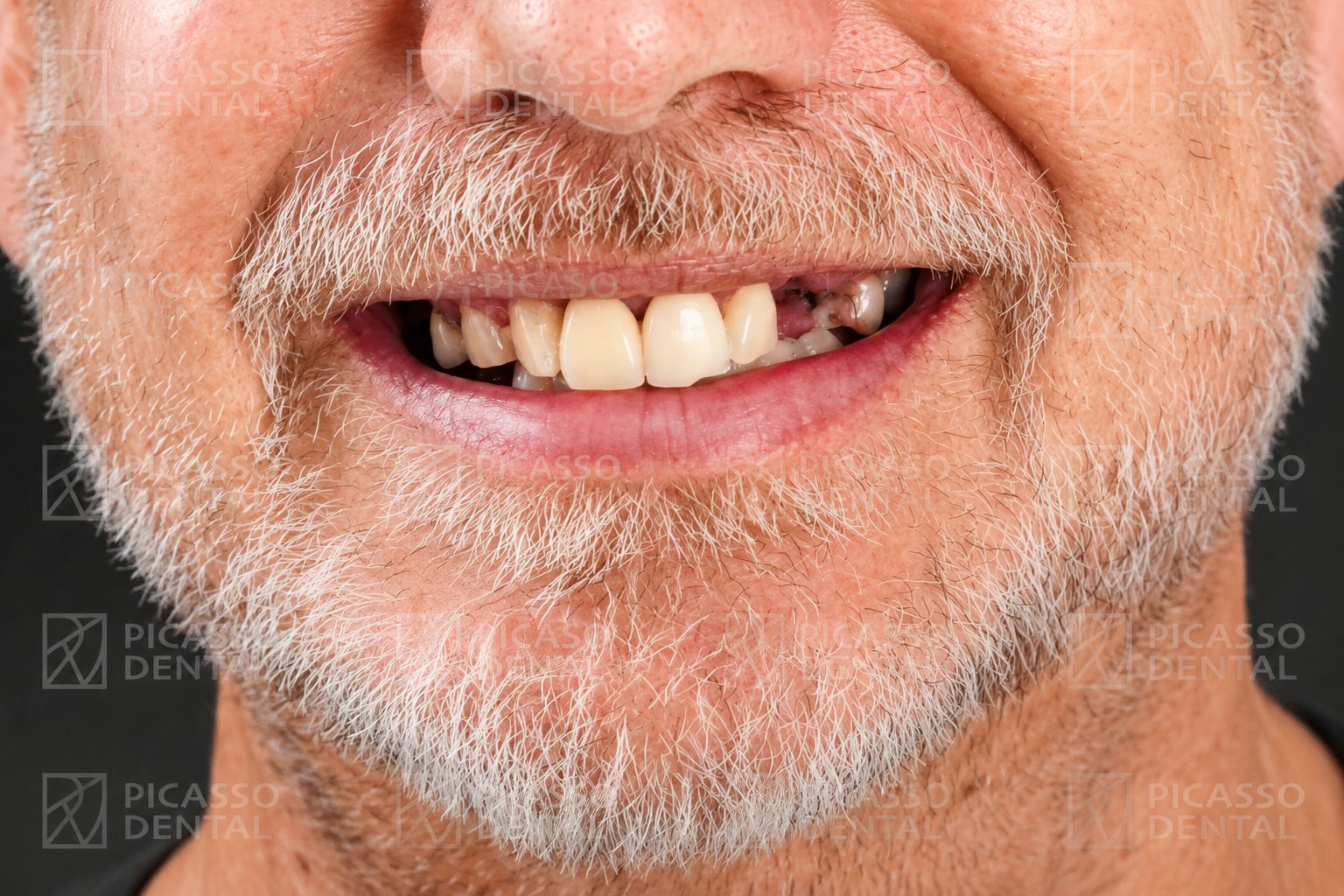 Before
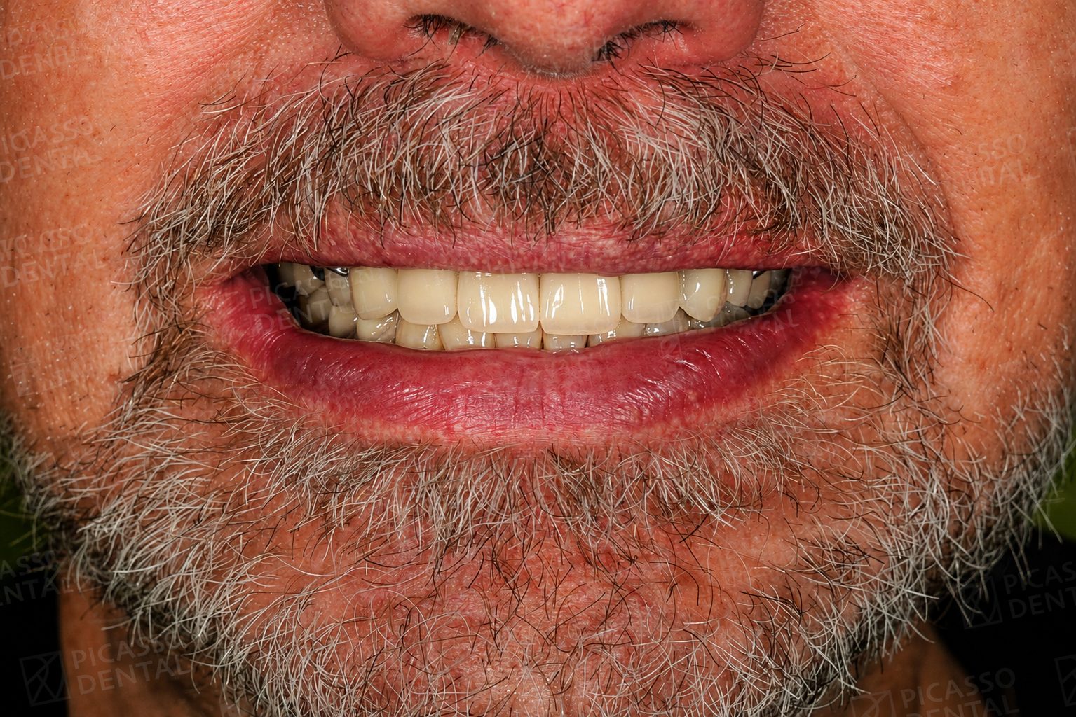 After
AfterCrown restoration, Hanoi Westlake
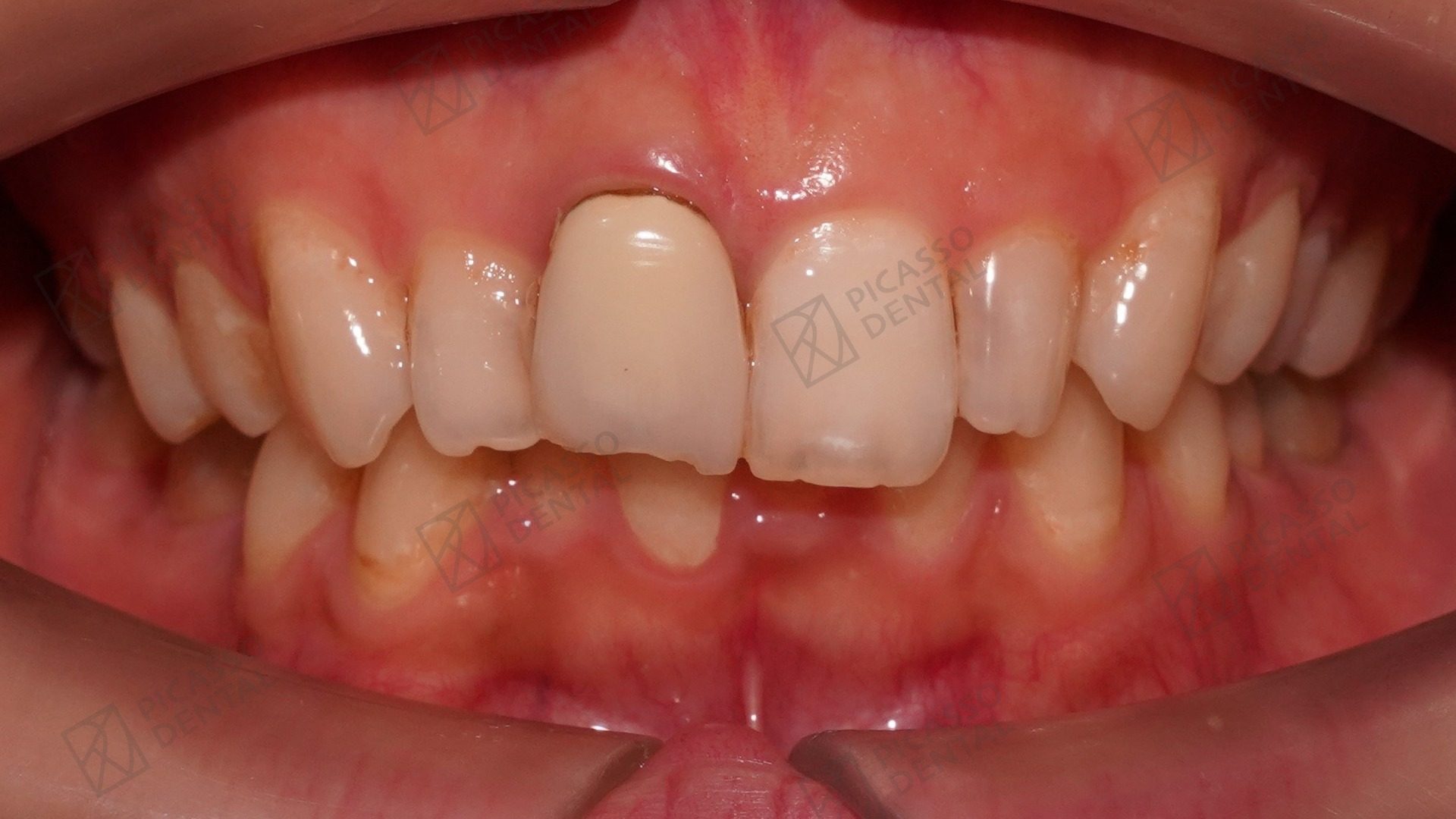 Before
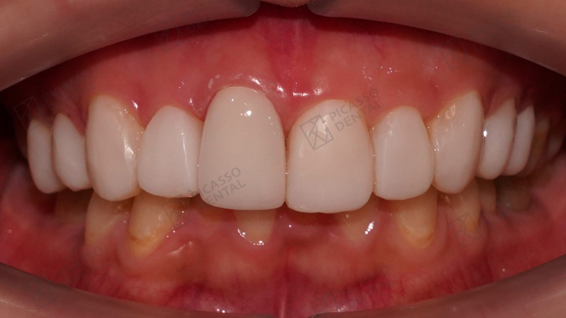 After
AfterSmile restoration, Da Nang Flagship
Frequently asked questions
What is a calcified root canal?
A calcified root canal is one where mineral deposits have narrowed or completely blocked the hollow space inside the tooth root that normally contains the nerve and blood vessels. This happens naturally with aging, as a protective response to trauma or chronic irritation, or due to untreated decay approaching the pulp. The canal space that should be hollow appears as solid tooth structure on X-rays, making standard root canal instruments unable to reach it.
Can a calcified root canal be treated?
In most cases, yes. Success rates for calcified root canal treatment range from 70 to 90 percent, lower than the 90 to 95 percent success rates for standard root canals. The outcome depends on the degree of calcification, the tooth type, and the experience and equipment of the treating dentist. Dental microscopes and ultrasonic instruments are essential for acceptable success rates. Without these tools, failure and perforation rates are significantly higher.
How is a calcified root canal different to treat than a standard root canal?
A standard root canal uses hand and rotary files to clean canals that are already accessible. A calcified canal requires locating the canal opening under high magnification, carefully removing the calcified material with ultrasonic instruments and EDTA chelating agent, and navigating slowly to avoid perforating the root wall. The procedure typically takes two to three times longer, requires two to three appointments, and demands specialist-level skill and equipment.
What equipment is required for calcified root canal treatment?
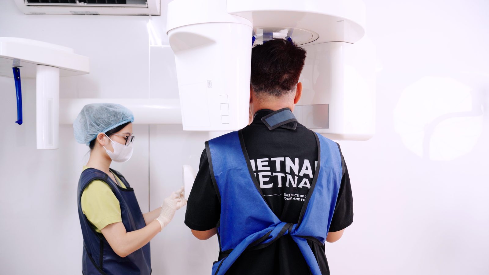
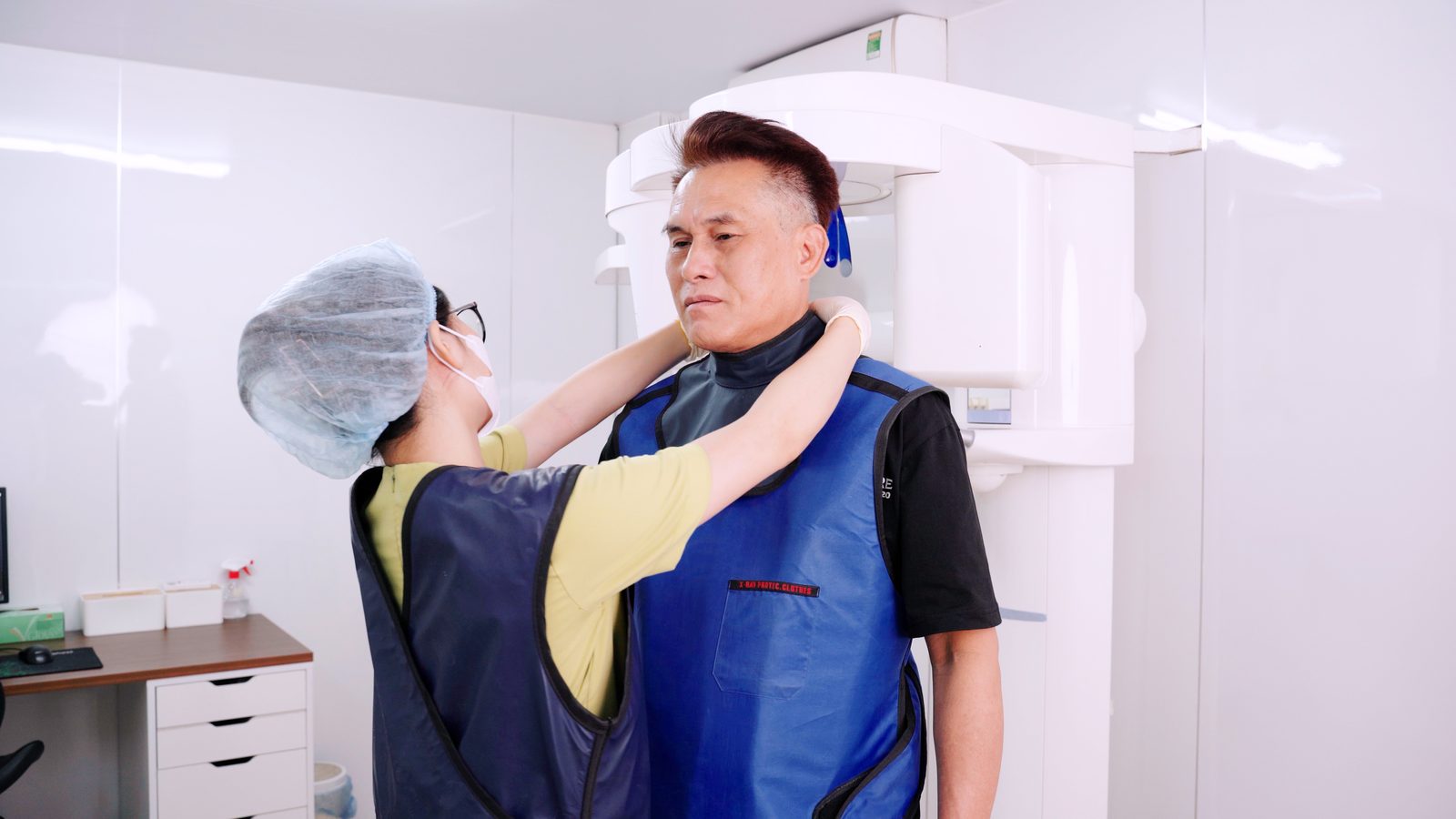
A dental microscope providing 8 to 20 times magnification is essential: without it, locating calcified canals is largely guesswork that leads to perforation. Ultrasonic instruments vibrate calcified material away without cutting tooth structure. EDTA chelating agent softens calcification chemically to facilitate removal. Cone beam CT scanning before treatment provides a three-dimensional map of canal anatomy that two-dimensional X-rays cannot reveal.
What happens if a calcified root canal cannot be treated?
When calcification is complete and canals cannot be accessed safely, two options remain. Apicoectomy (surgical root-end resection) removes the calcified root tip from outside the tooth and seals the canal from the bottom. If this is also not possible or fails, extraction followed by implant, bridge, or removable replacement is the remaining option. The decision between attempted treatment and immediate extraction should be made after CBCT assessment of the specific anatomy and an honest discussion of success probability.
How long does calcified root canal treatment take?
The first appointment typically takes two to three hours as the calcified canal is located, partial removal is performed, and medication is placed. A second appointment of 60 to 90 minutes completes the cleaning, shaping, and obturation. In complex cases a third appointment may be needed. Total treatment time is two to three times longer than a standard root canal on the same tooth.
Is calcified root canal treatment more expensive than standard root canal treatment?
Yes. The additional time, specialist equipment, and higher skill requirement mean calcified root canal treatment is priced above standard root canal fees at Picasso Dental Clinic. However, the total cost remains significantly less than equivalent treatment in Australia, the UK, or the United States, even when specialist endodontic care is included. A written quote is provided after CBCT assessment before any treatment begins.
How do I know if my tooth has a calcified canal?
Your dentist may suspect calcification when routine X-rays show no visible canal space inside the root, or when a tooth that should be sensitive shows no response to pulp testing. Cone beam CT scanning confirms the degree of calcification and maps the canal anatomy three-dimensionally. A history of dental trauma, prolonged grinding, or simply being over 50 all increase the likelihood of calcification in teeth requiring root canal treatment.
Start Here
Start with a brief.
Describe your tooth situation and a licensed endodontic dentist will reply with a written assessment of your treatment options and realistic success probabilities within twenty-four hours. No obligation.